
 Worksheets
A&P Section D exam review
Total questions: 136
Worksheet time: 2hrs 46mins
embolus dam
thrombus plug
platelet dam
embolus
platelet cork
BLOOD VESSELS that carry blood AWAY from heart
BLOOD VESSELS that carry blood back toward the heart
If the papillary muscles fail to contract,
Blood will not enter the atria, causing a heart attack
The ventricles will not pump blood, causing edema
The AV valves will not close properly, backflow of blood into atria occurs
The aortic and pulmonary valves will not open
Which part of the heart receives blood?
Atria
Aorta
Ventricles
Pulmonary Artery
Which type of blood vessels carry blood toward the heart?
Arteries
Venules
Arterioles
Capillaries
Which blood vessels are small and thin enough to allow for the diffusion of gases and nutrients into and out of circulation?
Capillaries
Venules
Arterioles
Veins
What side of the heart carries deoxygenated blood?
Left
Right
Both sides of the heart carry deoxygenated blood
Neither; blood is always oxygenated when it travels through the heart.

Identify the structure labeled at 1
Pulmonary Vein
Superior Vena Cava
Inferior Vena Cava
Pulmonary Artery

Identify the structure at 5
Aorta
Pulmonary Artery
Superior Vena Cava
Pulmonary Vein

Identify the structure at 8
Pulmonary Artery
Pulmonary Vein
Inferior Vena Cava
Aora

Identify the structure at 4
Superior Vena Cava
Pulmonary Artery
Inferior Vena Cava
Pulmonary Vein

Identify the structure at 6
Superior Vena Cava
Aorta
Pulmonary Vein
Pulmonary Artery

Identify the most muscular chamber of the heart that pumps oxygenated blood into systemic circulation.
2
3
6
9

What is the name of the valve between chambers 7 and 9?
Pulmonary Valve
atrioventricular valve
semilunar valve
tricuspid

Which number represents the right atrium?
3
2
6
9

Which location of the heart is associated with the presence of oxygenated blood?
4
8
6
2

Choose the correct sequence of blood flow
L. Atrium, Pulmonary Vein, R. Atrium, Aorta
R. Atrium, Pulmonary Vein, Aorta, Vena Cava
R. Ventricle, R. Atrium, Pulmonary Artery, Aorta
L. Ventricle, Vena Cava, L. Atrium, Aorta

__________ Must be strong and elastic in order to withstand higher blood pressure leaving the heart's ventricles.
Arteries
Capillaries
Veins
Venules
Which respiratory structure comes in direct contact with the circulatory system as Carbon Dioxide gas diffuses out of the body and Oxygen diffuses in?
diaphragm
bronchioles
Alveoli
bronchi

common carotid artery
subclavian artery
brachial artery
jugular artery

brachial vein
ulnar vein
cephalic vein
radial vein

axillary vein
cephalic vein
brachial vein
subclavian vein

jugular vein
axillary vein
iliac vein
radial vein

inferior vena cava
superior vena cava
renal vein
popliteal vein


ECG and EKG are abbreviations for the same thing
True
False
Resting period between heart contractions, when the heart fills with blood. Lower number of blood pressure.
Diastole
Systole
Venule
Arteriole
Period of contraction in the atria and ventricles. Larger number in blood pressure measurement based on this.
Systole
Diastole
Pulmonary refers to the
Lungs
Kidneys
Brain
Heart
Dividing tissue between the ventricles
Septum
Sacrum
Labrum
Larger of the two types of heart chambers
Ventricles
Atria

Perkinje fibers are found in the
Ventricles
Atria

Chordae Tendinae, or our heart strings, are attached to the
Valves
Arteries
Veins
SA and AV nodes
The right side of the heart (based on patient) pumps blood to the
Lungs
Body
Brain
Arms
The left side of the heart (based on patient left) pumps blood to the
Body
Brain
Lungs
The apex of the heart sits on this side of the chest, based on patient directions.
Left
Right
Atria and ventricles contract at the same time
True
False
Coronary arteries supply oxygenated blood to the
Heart
Lungs
Brain
Arms
When blood comes back from the body, it first flows into the
Right atria
Left atria
Right ventricle
Left ventricle
The first impulse for contraction originates in the
SA node
AV node
Perkinje fibers
Bundle of His
The electrical pathway shown on an ECG/EKG reading is described using
PQRST
QRS
ABCDE
12345
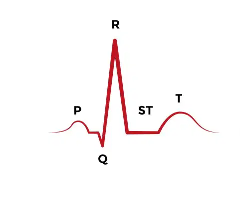
The largest peak and valley (QRS wave) represents
Ventricular contraction
Atrial contraction
Pacemaker of the heart, initiating contraction of right atria
SA node
AV node
Perkinje fibers
Septum
Where does a myocardial infarction occur?
Coronary arteries
Aorta
Septum
Pulmonary veins
What is the correct path through the circulatory system which describes the passage of blood originating in the left leg?
Vena cava → left atrium → left ventricle → lungs → right atrium → right ventricle → aorta
Vena cava → left atrium → right atrium → lungs → left ventricle → right ventricle → aorta
Vena cava → right atrium → right ventricle → lungs → left atrium → left ventricle → aorta
a) Vena cava → right atrium → left atrium → lungs → right ventricle → left ventricle → aorta
Which type of blood vessel has valves inside?
Artery
Vein
Capillary
None of the above
Which type of blood vessel has a 'pulse'?
Artery
Vein
Capillary
None of the above

Name the vessels labeled 1 and 9
Inferior Vena cava and Aorta
Inferior Vena cava and Left Pulmonary artery
Supeior Vena Cava and Left Pulmonary artery
Supeior Vena Cava and Right Pulmonary Artery
It allows the flow of blood in only one direction from the right atrium to the right ventricle. Which part of the human heart carries out this function?
Tricuspid valve
Bicuspid valve
Vena cave
Septum

Name part N.
Right atrium
left atrium
right ventricle
left ventricle

Name part M.
Tricuspid valve
Semilunar valve
Bicuspid valve
Mitral valve

The human circulatory system is called a double closed circulatory system. What are the 2 parts of the human circulatory system ?
The Pulmonary and Ventricular Circulation
The Pulmonary and Systemic Circulation
The Central and Peripheral Circulation
Small vessels that carry blood from arterioles and to small vessels called venules which then empty into veins
Arteries
Coronary Vessels
Valves
Capillaries
Structure around the heart
Transcardia
Circumcardia
Pericoronal
Pericardium
The smallest conducting respiratory passageways are called the
bronchioles
trachea
glottis
alveoli
Which of the following structures closes off the larynx during swallowing
glottis
epiglottis
esophagus
palate
The anatomical name for the windpipe is the
glottis
epiglottis
trachea
vocal cords
The main function of the squamous epithelial cells in the alveolar walls is
to create surfactant
stimulate the diaphragm
reduce surface tension
gas exchange
The main function of surfactant in the alveoli is
to reduce surface tension of the avleolar fluid
gas exchange
close off the larynx during swallowing
separate the oral and nasal cavities

Structure A represents
the epiglottis
the hyoid bone
thyroid cartilage
circoid cartilage

Structure B represents
the epiglottis
the hyoid bone
thyroid cartilage
circoid cartilage
All of the following are functions of the larynx EXCEPT
provides a patent airway
acts in voice production
serves as a switching mechanism to route food into the esophagus
stimulation of the diaphragm
alveoli
are specialized structures for gas exchange
have a large surface area for their size
are specialized for optimal diffusion
all of the previous answers are true of alveoli
1. larynx
2. nasal cavity
3. pharynx
4. trachea
Which of the following is the correct order that air would flow during inhalation.
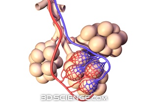
Responsible for sweeping mucus up out of the lungs.
alveoli
cilia
dermacytes
erythrocytes
These structures inside the nose help create turbulence in the incoming air, causing more contact with the mucus membrane.
Nasal Conchae
Paranasal Sinuses
Goblet Cells
Cilia
A bony layer that separates the nasal passages from the oral cavity, anteriorly.
Hard Palate
Soft Palate
Uvula
Epiglottis
Soft tissues that form the posterior portion of the separation between the nasal passages and oral cavity. Rises up to block nose while swallowing.
Soft Palate
Hard Palate
Surfactant
Pharynx

Structure indicated here with a scalpel (yikes!).
Uvula
Hard Palate
Palatine Tonsils
Epiglottis
The ___ lung is smaller because the ___ takes up some of its space.
Left, Heart
Left, Stomach
Right, Esophagus
Right, Right primary bronchus
The walls of the alveoli are composed of __ tissue.
Simple squamous
Simple cuboidal
Stratified squamous
Transitional epithelial
Made by the type II cells, this oily substance helps break the surface tension of moisture in the lungs, keeping the alveolar walls from sticking.
Surfactant
Mucus
Goblet cells
Niacinamide
What separates the parietal and visceral pleurae are separated by
Water
Air
Pleural fluid
Fats
What muscles are used in breathing in and breathing out?
Diaphragm and intercostals
Diaphragm and lungs
Lungs and ribs
Lungs and diaphragm
What is the volume of one breath called?
Minute ventilation
Inspiratory capacity
Tidal volume
Vital capacity
During inspiration, the intrapleural pressure mostly
Decreases
Increases
Fluctuates rapidly
Stays the same
Which diagram best illustrates the function of an alveolus?


What is the term for the tiny air sacs of the lungs which allow for rapid gas exchange?
bronchus
trachea
alveoli
bronchioles
The two branches off of the trachea are the left and right:
lungs
bronchioles
bronchus
diaphragm
What is the name of the tiny hairs that protect the nasal passageways, trachea and and other parts of the respiratory tract and filter out foreign matter and organisms that are inhaled with the air?
capillaries
bronchioles
cilia
alveoli
Which organ is also known as the voicebox?
nasal passage
larynx
trachea
bronchi

The "windpipe" is known as the
alveoli
esophagus
trachea
bronchi
A small flap of tissue that covers the trachea is called the
bronchi
larynx
pharynx
epiglottis
Which properly describes inhalation?
ribs go down and out
pressure is low causing air to rush in
pressure is high causing air to rush in
volume is low
Med term- Which med term means a deficiency of something?
-emia
-haem
-penia
-hyper
Med term- Which term means breath or breathing?
-pulm(o)
-pnea
bronch(o)
oxy
Med term- Please select ALL of the med terms that mean 'lung'.
pneum(o)
pneumon(o)
pulm(o)
thorac(o)

