Wayground logo

Free Printable Worksheets

Font size

S
M
L
XL
Worksheets

Unit 1.1 TEST REVIEW HBS

Total questions: 136

Worksheet time: 1hrs 17mins

Name
Class
Date
1.

What type of fracture is this? (Hint: it's across the bone.)

a)

Dorsal

b)

Linear

c)

Closed

d)

Transverse

2.

If a fracture is comminuted, that means the bone is in _______ pieces.

a)

Multiple

b)

One

3.

What type of fracture is this?

a)

Transverse

b)

Comminuted

c)

Simple

d)

Spiral

4.

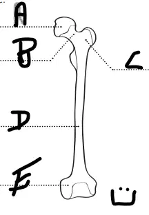
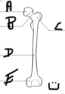
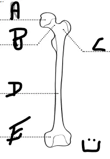
Where are the femoral condyles?

(a)  

5.

Where is the femoral lateral condyle?

(a)  

6.

Where is the femoral median condyle?

(a)  

7.

Where is the femoral shaft?

(a)  

8.

Where is the femoral head?

(a)  

9.

Where is the femoral neck?

(a)  

10.

Cells that can build bony matrix.

a)

Osteoblasts

b)

Osteoclasts

c)

Canaliculi

d)

Lamellae

11.

Cells that dissolve and reabsorb the bony matrix.

a)

Osteoclasts

b)

Osteoblasts

c)

Canaliculi

d)

Lamellae

12.

Bones are considered alive.

a)

True

b)

False

13.

What is the blood clot that first forms around a fracture?

a)

Hematoma

b)

Callus

c)

Bone

14.

Which type of fracture is demonstrated in this image?

a)

Simple

b)

Spiral

c)

Comminuted

d)

Compound

15.

Name the fracture

a)

transverse femur fracture

b)

spiral humerus fracture

c)

transverse humerus fracture

16.

Name the fracture

a)

transverse femur fracture

b)

comminuted femur fracture

17.

First step of fracture healing is the formation of ___________.

a)

Hematoma

b)

Fibrocartilage callus

c)

Epiphyseal line

d)

Bony callus

18.

What occurs in the third step of fracture healing?

a)

Fibrocartilaginous converts to bony callus

b)

Bone remodeling

c)

Torn blood vessels hemorrhage

d)

Compact bone is laid down

19.

In the fourth step of fracture healing this will occur

a)

Fibrocartilaginous converts to bony callus

b)

Bony callus formation

c)

Bone remodeling

d)

Mass or clot is forming

20.

What is happening in this stage of bone healing?

a)

Bone is fractured, hematoma is formed

b)

Bony callus forms, osteoblast cells create new bone

c)

Remodeling, osteoclasts remove extra bone until its completely healed

d)

Fibrocartilage callus forms, chondroblast and fibroblasts produce the fibrocartilaginous calus

21.

What is happening in this stage of bone healing?

a)

Bone is fractured, hematoma forms

b)

Bony callus forms, osteoblast cells create new bone

c)

Remodeling, osteoclasts remove extra bone until its completely healed

d)

Fibrocartilage callus forms, chondroblast and fibroblasts produce the fibrocartilaginous calus

22.

What is happening in this stage of bone healing?

a)

Bone is fractured

b)

Bony callus forms, osteoblast cells create new bone

c)

Remodeling, osteoclasts remove extra bone until its completely healed

d)

Connective tissue cells form a fibrocartilage callus . New capillaries begin to form in the damaged area.

23.

Place the following stages of fracture healing in the correct order

a)

Hematoma Formation

b)

Fibrocartilage Callus Formation

c)

Bony Callus Formation

d)

Bone Remodeling

1)
2)
3)
4)
24.

Which of the following scans does not use radiation?

a)

MRI

b)

CT scan

c)

PET scan

d)

X-ray

25.

Ultrasound is a safe procedure that uses ______________.

a)

radiation

b)

sound waves

c)

dye

d)

film

26.
Which of these is a bone cell?
a)
osteoclast
b)
osteoblast
c)
osteoma
d)
ostecoma
27.
What is the first stage of fracture healing?
a)
hematoma formation
b)
fibrocartilage callus formation
c)
bony callus formation
d)
bone remodeling
28.
What is the third stage of fracture healing?
a)
hematoma formation
b)
fibrocartilage callus formation
c)
bony callus formation
d)
bone remodeling
29.
What would be the best label for line 'A'?
a)
femoral head
b)
femoral neck
c)
shaft
d)
condyles
e)
trochanteric region
30.
What would be the best label for line 'C"?
a)
trochanteric region
b)
femoral head
c)
femoral neck
d)
shaft
e)
condyles
31.
What would be the best label for line 'E"?
a)
condyles
b)
femoral head
c)
femoral neck
d)
shaft
e)
trochanteric region
32.
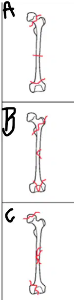
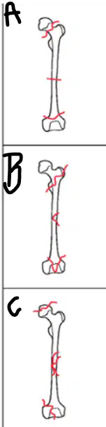
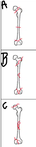
Which of these shows a 'transverse, extra-articular, femoral neck' fracture?
a)
A
b)
B
c)
C
33.
Which of these shows a comminuted, intra-articular, femoral head fracture?
a)
A
b)
B
c)
C
34.
Which of these shows a spiral, intra-articular, femoral trochanteral region fracture?
a)
A
b)
B
c)
C
35.
#9
a)
zygomatic bone
b)
parietal bone
c)
occipital bone
d)
frontal bone
e)
mandible
36.
Spinal cord & brain - send & receive signals
a)
Nervous Tissue
b)
Muscle Tissue
c)
Epithelial Tissue
d)
Connective Tissue
37.
#3
a)
occipital bone
b)
mandible
c)
nasal bone
d)
temporal bone
e)
maxilla
38.
#9
a)
zygomatic bone
b)
parietal bone
c)
occipital bone
d)
frontal bone
e)
mandible
39.
Flat muscles on the sides of the head that connect to the mandible and allow it to move and chew.
a)
Orbicularis oris
b)
Temporalis
c)
Frontalis
d)
Orbicularis oculi
40.
Circular muscle around the mouth that allows the lips to move.
a)
Temporalis
b)
Orbicularis oris
c)
Orbicularis oculi
d)
Frontalis
41.
The muscle directly over the frontal bone that allows us elevate the eyebrows
a)
Orbicularis oculi
b)
Orbicularis oris
c)
Temporalis
d)
Frontalis
42.
Cells that work together to perform a similar function
a)
organs
b)
organisms
c)
organ systems
d)
tissues
43.
Profesionals who study the microscopic anatomy of tissues
a)
EMTs
b)
histologists
c)
endocrinologist
d)
herpatologists
44.
#6
a)
temporal bone
b)
parietal bone
c)
zygomatic bone
d)
frontal bone
e)
occipital bone
45.
(1.2.2) How many thoracic vertebrae are there?
a)
5
b)
27
c)
7
d)
12
46.
During testing of our cohort patients, one patient’s tissue showed evidence of abnormal tissue.  Testing confirmed he/she had carncer of glandular epithelial cells.  Which patient?
a)
Hazel
b)
Prue
c)
Dillon
d)
Jackson
e)
Ren
47.
Found where tissues connect (i.e. between muscles/bones and bones/joints) - supports/connects other tissue types
a)
Nervous Tissue
b)
Muscle Tissue
c)
Epithelial Tissue
d)
Connective Tissue
48.
A circular muscle around the eye socket. These muscles open and close the eyelids.
a)
Frontalis
b)
Orbicularis oculi
c)
Orbicularis oris
d)
Temporalis
49.
#5
a)
maxilla
b)
parietal bone
c)
temporal bone
d)
occipital bone
e)
mandible
50.
Striated/skeletal, smooth (walls of organs) or cardiac (in heart)
a)
Nervous Tissue
b)
Muscle Tissue
c)
Epithelial Tissue
d)
Connective Tissue
51.
Spinal cord & brain - send & receive signals
a)
Nervous Tissue
b)
Muscle Tissue
c)
Epithelial Tissue
d)
Connective Tissue
52.
Tissues that work together to perform a similar function
a)
tissues
b)
organisms
c)
organ systems
d)
organs
53.
#4
a)
mandible
b)
parietal bone
c)
occipital bone
d)
maxilla
e)
zygomatic bone
54.
#2
a)
mandible
b)
zygomatic bone
c)
temporal bone
d)
occipital bone
e)
frontal bone
55.
Lining surfaces of organs & blood vessels - absorbs, secretes, protects & senses
a)
Nervous Tissue
b)
Muscle Tissue
c)
Epithelial Tissue
d)
Connective Tissue
56.
The way in which a living organism or bodily part functions.
a)
Organ systems
b)
Physiology
c)
Histology
d)
Pathology
57.
#7
a)
temporal bone
b)
frontal bone
c)
nasal bone
d)
parietal bone
e)
occipital bone
58.
#10
a)
parietal bone
b)
maxilla
c)
nasal bone
d)
zygomatic bone
e)
temporal bone
59.
The bone most anterior if this patient were laying on their back
a)
Femur
b)
Fibula
c)
Patella
d)
Tibia
60.
The hamstrings are anterior or posterior?
a)
anterior
b)
posterior
61.

The hands are ____ to the feet.

a)

superior

b)

inferior

c)

superficial

d)

deep

62.

The heel is ___ to the toes.

a)

anterior

b)

deep

c)

posterior

d)

intermediate

63.

The knee is _____ to the thigh.

a)

distal

b)

proximal

c)

medial

d)

lateral

64.

The heart is medial to the arm.

a)

TRUE

b)

FALSE

65.

The skin is superficial to the skeletal muscles.

a)

TRUE

b)

FALSE

66.

toward side of the body

a)

ventral

b)

dorsal

c)

medial

d)

lateral

67.

Which anatomical plane is the ONLY horizontal plane?

a)

sagittal

b)

transverse

c)

frontal

68.

Which anatomical plane divides the body into anterior and posterior

a)

frontal

b)

transverse

c)

sagittal

d)

midsagittal

69.

Plane that divides body into left and right sides:

a)

Sagittal

b)

medial

c)

transverse

70.
This type of tissue lines the internal organs and covers the outside of the body as  skin
a)
epithelial
b)
connective
c)
muscular
d)
nervous
71.

This type of tissue relays messages

a)

epithelial

b)

connective

c)

muscular

d)

nervous

72.

What type of tissue is this?

a)

connective tissue

b)

nervous tissue

c)

epithelial tissue

d)

muscle tissue

73.

What type of tissue is this?

a)

epithelial

b)

muscle

c)

nervous

d)

connective

74.

What type of tissue is this?

a)

nervous tissue

b)

muscle tissue

c)

epithelial tissue

d)

connective tissue

75.
What type of bone is the femur?
a)
Long bone
b)
Short bone
c)
Flat bone
d)
Irregular bone
76.
Name the bone located at number  8.
a)
Humerus
b)
Radius
c)
Fibula
d)
Tibia
77.
The shaft of the long bone. 
a)
epphysis
b)
diaphysis
c)
epiphyseal line
d)
growth plate
78.
The shiny white substance at #8 is the 
a)
articular cartilage
b)
periosteum
c)
yellow marrow
d)
diaphysis
79.
In adults, the substance at #6 would be 
a)
red marrow
b)
yellow marrow
c)
spongy bone
d)
clotted blood
80.
The epiphysis is 
a)
The shaft of a bone
b)
a bone cell
c)
the end of a long bone
d)
the covering of a bone
81.

The only freely moveable bone in the skull is the ______.

a)

frontal

b)

maxilla

c)

occipital

d)

mandible

82.

How many vertebrae are in the thoracic region?

a)

12

b)

20

c)

10

d)

7

83.

How many vertebrae are in the neck?

a)

2

b)

4

c)

7

d)

12

84.

Which is the correct order of ribs from superior to inferior?

a)

floating, true, false

b)

true, false, floating

c)

true, floating, false

d)

false, floating, true

85.
What is the name of this bone?
a)
Femur
b)
Humerous
c)
Skull
d)
Shin
86.

Identify bone "F"

a)

Temporal

b)

Parietal

c)

Sphenoid

d)

Zygomatic

87.

Identify Bone "H"

a)

Zygomatic

b)

Maxilla

c)

Sphenoid

d)

Lacrimal

88.

identify Bone "G"

a)

Maxilla

b)

Mandible

c)

Zygomatic

d)

Vomer

89.
a)
Zygomatic
b)
Temporal
c)
Parietal
d)
Lacrimal
90.
a)
Zygomatic
b)
Temporal
c)
Parietal
d)
Occipital 
91.
a)
Zygomatic
b)
Frontal
c)
Parietal
d)
Lacrimal
92.
Large bone; makes up the roof of the orbital and nasal cavities; "forehead". 
a)
frontal 
b)
sphenoid 
c)
parietal 
d)
ethmoid 
93.
a)

frontal

b)

sphenoid

c)

zygomatic

d)

nasal

94.
a)

maxilla

b)

mandible

c)

zygomatic

d)

parietal

95.
two bones, side-by-side that make up the bridge of the nose; 
a)
sphenoid bones
b)
zygomatic bones 
c)
lacrimal bones 
d)
nasal bones 
96.
a)
Occipital
b)
Parietal
c)
Temporal
d)
Sphenoid
97.
#3
a)
occipital bone
b)
mandible
c)
nasal bone
d)
temporal bone
e)
maxilla
98.
#9
a)
zygomatic bone
b)
parietal bone
c)
occipital bone
d)
frontal bone
e)
mandible
99.
Flat muscles on the sides of the head that connect to the mandible and allow it to move and chew.
a)
Orbicularis oris
b)
Temporalis
c)
Frontalis
d)
Orbicularis oculi
100.
Circular muscle around the mouth that allows the lips to move.
a)
Temporalis
b)
Orbicularis oris
c)
Orbicularis oculi
d)
Frontalis
101.
The muscle directly over the frontal bone that allows us elevate the eyebrows
a)
Orbicularis oculi
b)
Orbicularis oris
c)
Temporalis
d)
Frontalis
102.
Cells that work together to perform a similar function
a)
organs
b)
organisms
c)
organ systems
d)
tissues
103.
Profesionals who study the microscopic anatomy of tissues
a)
EMTs
b)
histologists
c)
endocrinologist
d)
herpatologists
104.
#6
a)
temporal bone
b)
parietal bone
c)
zygomatic bone
d)
frontal bone
e)
occipital bone
105.
(1.2.2) How many thoracic vertebrae are there?
a)
5
b)
27
c)
7
d)
12
106.
During testing of our cohort patients, one patient’s tissue showed evidence of abnormal tissue.  Testing confirmed he/she had carncer of glandular epithelial cells.  Which patient?
a)
Hazel
b)
Prue
c)
Dillon
d)
Jackson
e)
Ren
107.
Found where tissues connect (i.e. between muscles/bones and bones/joints) - supports/connects other tissue types
a)
Nervous Tissue
b)
Muscle Tissue
c)
Epithelial Tissue
d)
Connective Tissue
108.
A circular muscle around the eye socket. These muscles open and close the eyelids.
a)
Frontalis
b)
Orbicularis oculi
c)
Orbicularis oris
d)
Temporalis
109.
#5
a)
maxilla
b)
parietal bone
c)
temporal bone
d)
occipital bone
e)
mandible
110.
Striated/skeletal, smooth (walls of organs) or cardiac (in heart)
a)
Nervous Tissue
b)
Muscle Tissue
c)
Epithelial Tissue
d)
Connective Tissue
111.
Spinal cord & brain - send & receive signals
a)
Nervous Tissue
b)
Muscle Tissue
c)
Epithelial Tissue
d)
Connective Tissue
112.
Tissues that work together to perform a similar function
a)
tissues
b)
organisms
c)
organ systems
d)
organs
113.
#4
a)
mandible
b)
parietal bone
c)
occipital bone
d)
maxilla
e)
zygomatic bone
114.
#2
a)
mandible
b)
zygomatic bone
c)
temporal bone
d)
occipital bone
e)
frontal bone
115.
Lining surfaces of organs & blood vessels - absorbs, secretes, protects & senses
a)
Nervous Tissue
b)
Muscle Tissue
c)
Epithelial Tissue
d)
Connective Tissue
116.
The way in which a living organism or bodily part functions.
a)
Organ systems
b)
Physiology
c)
Histology
d)
Pathology
117.
#7
a)
temporal bone
b)
frontal bone
c)
nasal bone
d)
parietal bone
e)
occipital bone
118.
#10
a)
parietal bone
b)
maxilla
c)
nasal bone
d)
zygomatic bone
e)
temporal bone
119.

What type of tissue is in this image?

a)

epithelial

b)

cartilage

c)

bone

d)

muscle

120.

What type of tissue is in this image

a)

Connective tissue

b)

Muscle tissue

c)

Epithelial tissue

d)

Nervous tissue

121.

What type of tissue is in this image?

a)

Areolar tissue

b)

Adipose tissue

c)

Dense connective tissue

d)

Bone tissue

122.

What type of tissue is in this image?

a)

connective tissue

b)

Bone tissue

c)

Adipose tissue

d)

Blood tissue

123.

What type of tissue is in this image?

a)

Adipose tissue

b)

Blood tissue

c)

Bone tissue

d)

Areolar tissue

124.

What type of tissue is in this image?

a)

Elastic

b)

Blood tissue

c)

Adipose tissue

d)

Areolar tissue

125.

What type of muscle tissue is in this image?

a)

Skeletal muscle tissue

b)

Cardiac muscle tissue

c)

Smooth muscle tissue

d)

Love muscle tissue

126.

What type of muscle tissue is in this image?

a)

Skeletal muscle tissue

b)

Cardiac muscle tissue

c)

Smooth muscle tissue

d)

Bicep muscle tissue

127.

What type of muscle tissue is in this image?

a)

Skeletal muscle tissue

b)

Cardiac muscle tissue

c)

Nervous muscle tissue

d)

Smooth muscle tissue

128.

What type of nervous tissue is this pictured in yellow?

a)

Neuroglial nervous tissue

b)

Neuron nervous tissue

c)

Smooth nervous tissue

d)

Cardiac nervous tissue

129.

Which is NOT one of the four types of tissues?

a)

Skeletal

b)

Connective

c)

Nervous

d)

Epithelial

130.

What type of tissue is this?

a)

Bone

b)

Connective

c)

Nervous

d)

Muscle

131.

a)

Reticular

b)

Fribrocartilage

c)

Elastic

d)

Nervous

132.

The study of tissues

a)

Cytology

b)

Histology

c)

Anatomy

d)

Physiology

133.

What type of epithelial tissue is this?

a)

Simple squamous

b)

simple columnar

c)

simple ciliated columnar

d)

simple cuboidal

134.

Bone and blood are examples of what type of tissue?

a)

Connective

b)

Epithelial

c)

Muscle

d)

Nervous

135.

What type of tissue is this?

a)

epithelial

b)

connective

c)

nervous

d)

muscle

136.

This type of tissue contracts and has lots of mitochondria

a)

epithelial

b)

connective

c)

nervous

d)

muscle