
 Worksheets
Chapter 10 Muscles Review
Total questions: 115
Worksheet time: 1hrs 3mins
Which of the following is the primary function of the muscular system?
Digestion
Body movement
Hormone production
Blood circulation
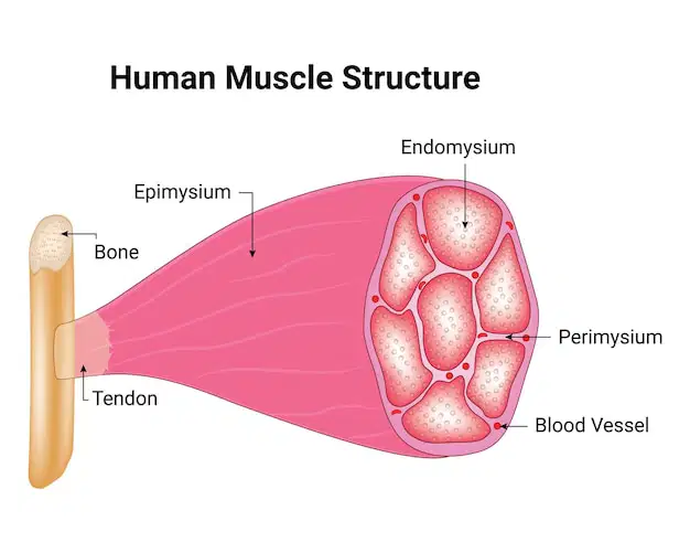
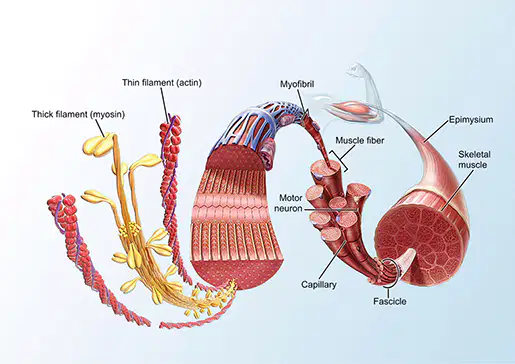
Skeletal Muscles: Focus on skeletal muscles that move bones, interact with the skeletal system in a lever mechanism, and are composed of muscle fibers and connective tissue layers:
Wraps individual fibers.
Endomysium
Groups fibers into fascicles.
Perimysium
Encases the entire muscle.
Epimysium
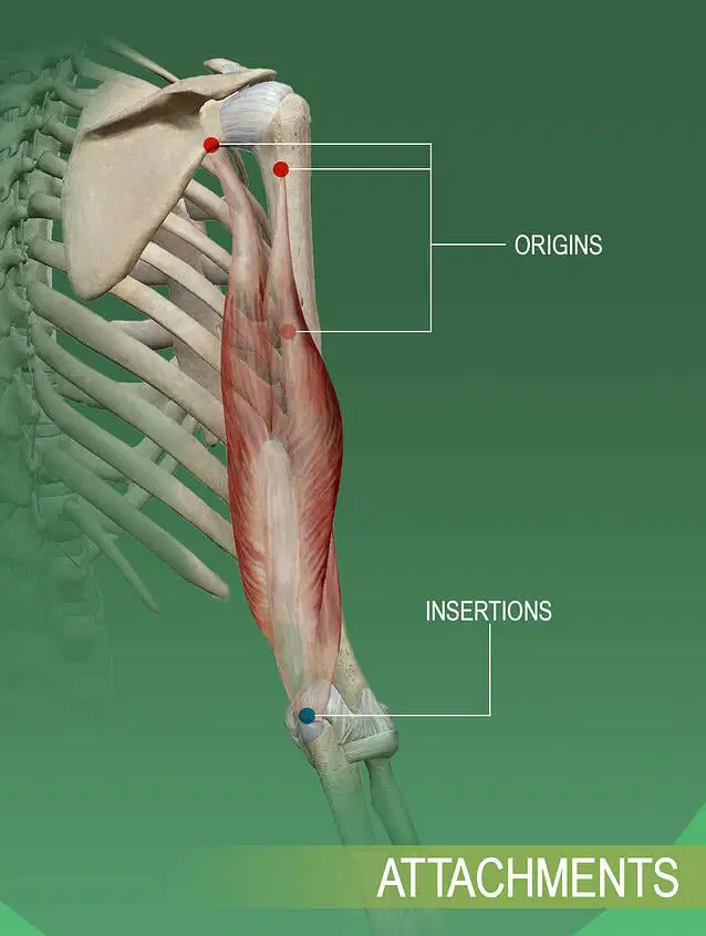
Muscle Attachments:
Origins and Insertions:
Stationary point of attachment.
Origin
Movable point of attachment.
Insertion
(extra space)
put me on the extra space!
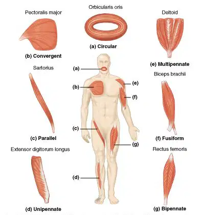
Fascicle Arrangements:
Side-by-side arrangement.
Parallel
Tapered ends with a bulging center.
Fusiform
Broad origin converging to a single insertion.
Convergent
Arranged in concentric rings.
Circular
Oblique arrangement to a central tendon (unipennate or bipennate).
Pennate
Muscle Interaction:
Main muscle responsible for a movement.
Prime Mover
Muscles opposing the movement.
Antagonists
Assist the prime mover, with fixators stabilizing joints.
Synergists

which skeletal muscle is represented in the image?
brachioradialis
deltoideus
biceps brachii
biceps femoris

which skeletal muscle is represented in the image?
brachioradialis
biceps brachii
biceps femoris
deltoideus

which skeletal muscle is represented in the image?
biceps femoris
semimembranosus
semitendinosis
gastrocnemius

which skeletal muscle is represented in the image?
deltoideus
triceps brachii
infraspinatus
teres major

which skeletal muscle is represented in the image?
depressor anguli oris
orbicularis oris
zygomaticus
sternocleidomastoid

which skeletal muscle is represented in the image?
external obliques
serratus anterior
rectus abdominis
pectoralis major

which skeletal muscle is represented in the image?
fibularis longus
tibialis anterior
gastrocnemius
sartorius

which skeletal muscle is represented in the image?
frontalis
orbicularis oculi
orbicularis oris
depressor anguli oris

which skeletal muscle is represented in the image?
gastrocnemius
tibialis anterior
fibularis longus
vastus medialis

which skeletal muscle is represented in the image?
gluteus medius
gluteus maximus
semitendinosus
external obliques

which skeletal muscle is represented in the image?
gluteus medius
gluteus maximus
semitendinosus
external obliques

which skeletal muscle is represented in the image?
latissmus dorsi
teres major
infraspinatus
deltoideus

which skeletal muscle is represented in the image?
orbicularis oculi
zygomaticus
orbicularis oris
frontalis

which skeletal muscle is represented in the image?
pectoralis major
serratus anterior
rectus abdominis
deltoideus

which skeletal muscle is represented in the image?
pectoralis major
serratus anterior
rectus abdominis
deltoideus

which skeletal muscle is represented in the image?
rectus femoris
vastus lateralis
vastus medialis
sartorius

which skeletal muscle is represented in the image?
semitendinosus
semimembranosus
biceps femoris
gastrocnemius

which skeletal muscle is represented in the image?
semitendinosus
semimembranosus
biceps femoris
gastrocnemius

which skeletal muscle is represented in the image?
serratus anterior
rectus abdominis
external obliques
pectoralis major

which skeletal muscle is represented in the image?
sternocleidomastoid
trapezius
zygomaticus
depressor anguli oris

which skeletal muscle is represented in the image?
sternocleidomastoid
trapezius
zygomaticus
depressor anguli oris

which skeletal muscle is represented in the image?
sternocleidomastoid
trapezius
zygomaticus
depressor anguli oris

which skeletal muscle is represented in the image?
triceps brachii
latissimus dorsi
deltoideus
brachioradialis
Which of the following is NOT true about oxygen debt?
During exertion, oxygen has been depleted
In recovery, we owe the body some oxygen.
Oxygen demands remain elevated in order to reduce high levels of lactic acid
This is the biggest reason someone passes out after exerting a lot of energy?.
These projections, or myosin heads, that are found on the thick filaments are called _____.
cross bridges
Z lines
A bands
actin tails
What happens during the power stroke of muscle contraction?
break down of ATP occurs at the myosin head so that it is ready to pull at actin again
cross bridges attach to actin filaments and pull them toward the middle of the sarcomere
when a muscle is unable to contract even through it is still being stimulated
oxygen debt
muscle fatigue
motor units
System that provides channels for ion flow throughout the muscle fibers to facilitate the propagation of an action potential.
transverse tubules
motor units
sarcoemeres
sarcoplasmic reticulum
A motor neuron and all of the muscle fibers it innervates/ controls
transverse tubules
motor units
sarcoemeres
sarcoplasmic reticulum
What ions free the actin strands and allow muscle contraction to occur?
POTASSIUM
CALCIUM
SODIUM
What energy molecule binds to the myosin heads and allows the detachment from actin?
GLUCOSE
ADP
ATP
CALCIUM
Microfilaments of actin and myocin in repeating subunits.
MYOFIBRILS
Z LINES
SARCOMERES
SARCOLEMMA
The contractile unit of a myofibril consisting of Z-lines, A-bands, and I-bands.
A BANDS
Z LINES
SARCOMERES
SARCOLEMMA
point of contact between a motor neuron and a skeletal muscle cell
I band
neuromuscular junction
Sarcoplasmic reticulum
Epimysium
A structure within a unit of myofibril. It is a section of the myofibril which contains myosin filaments.
A band
I bands
Z line
sarcomere
A structure within a unit of myofibril. It is a band which contains only actin fibers.
A band
I bands
Z line
sarcomere
A structure within a unit of myofibril. It is a membrane which actin is embedded in.
A band
I bands
Z line
sarcomere
Neurotransmitter that produces an impulse in a muscle cell
serotnoin
dopamine
epinephrine
acetylcholine
Which of the following is NOT a protein found in every myofibril?
ACTIN
MYOSIN
HISTONE
What does the sliding filament model strive to explain?
OXYGEN DEBT
MUSCLE FATIGUE
MUSCLE CONTRACTION
What two proteins prevent the myosin heads from binding to the actin strands?
troponin
globulins
tropomyosin
fibrin
What do you call the microfilaments of actin and myocin in repeating subunits?
myosin
myofibril
Which of the following is NOT an pathway the skeletal muscles use for ATP regeneration?
CELLULAR (Aerobic) respiration
Breaking down ADP to produce ATP faster
Anaerobic respiration
Direct phosphorylation of ADP by creatine phosphate

The epimysium covers
the entire muscle
the fascicle
the muscle fiber
the myofibril

Which label is the muscle fiber?
A
B
C
D

Which label is the myofibril?
A
B
C
D

What is another name for #9 (cell membrane)?
Sarcomere
Sarcolemma
Myofibril
Myofiber
Which type of muscle tissue is found in the heart?
Skeletal muscle tissue
Smooth muscle tissue
Cardiac muscle tissue
None of the above
Which type of muscle tissue is found in the digestive system?
Skeletal muscle tissue
Smooth muscle tissue
Cardiac muscle tissue
None of the above
Which type of muscle tissue is involved in voluntary control?
Skeletal muscle tissue
Smooth muscle tissue
Cardiac muscle tissue
None of the above
What is the function of intercalated discs in cardiac muscle tissue?
To aid in muscle contraction
To provide elasticity
To allow conscious control
None of the above
What is the function of smooth muscle tissue in the bladder?
To aid in muscle contraction
To provide elasticity
To allow conscious control
None of the above
What is the main muscle doing the work called?
Agonist
Antagonist
Origin
Insertion
What is the term for the ability of muscle tissue to be stimulated?
Extensibility
Elasticity
Excitability
Contractility
What is the protein that makes up thin filaments in a sarcomere?
Actin
Myosin
Tropomyosin
Troponin
What is the protein that makes up thick filaments in a sarcomere?
Actin
Myosin
Tropomyosin
Troponin
What is the role of calcium in muscle contraction?
It binds to troponin and allows myosin to bind to actin
It hydrolyzes ATP to provide energy for muscle contraction
It blocks the myosin bonding sites on actin
It causes the thin and thick filaments to slide past each other

Long ribbon-like contractile organelles that take up most of the cell.
nuclei
mitochondria
T- tubules
myofibrils
myosin

Thick myofilament made of protein with cross bridges
nuclei
mitochondria
myofibrils
myosin

Plasma membrane in muscle cell
sarcolemma
sarcoplasma
sarcoplasmic reticulum
sarcomere
Fluid part of a muscle cell (fiber)
sarcolemma
sarcoplasma
sarcoplasmic reticulum
sarcomere

Series of tubes that store calcium ion
sarcolemma
sarcoplasma
sarcoplasmic reticulum
sarcomere

Contractile unit in myofibrils
sarcolemma
sarcoplasma
sarcoplasmic reticulum
sarcomere

Thin myofilament made of contractile protein
sarcolemma
myosin
sarcomere
actin

Controls the cell's activity. More than one found in muscle cell/fiber
nuclei
mitochondria
T- tubules
myosin

An individual muscle cell (7) is called a ...
myofibril
fascicle
sarcomere
fiber

Individual muscle cells are encased in a tough connective tissue sheath called the ...
sarcoplasmic reticulum
endomysium
perimysium
epimysium

Muscle cells are bundled together to form (6) ...
fascia
myofibrils
sarcomeres
fascilcles

Bundles of muscle cells are bound together by connective tissue (5) called ...
epimysium
perimysium
sarcomysium
endomysium

An entire muscle is wrapped in a tough translucent sheath called the (3) ...
endomysium
fascia
perimysium
sarcomysium

Muscles attach to bones by (1) ...
tendons
ligaments
sarcomeres
myofibrils
Because of their energy requirements, muscle cells have an unusually large number of ...
vacuoles
ribosomes
nuclei
mitochondria
Which type of muscle is striated?
skeletal
smooth
cardiac
Which connective tissue surrounds the fascicle?
epimysium
perimysium
sarcolemma
endomysium
The epimysium covers
the entire muscle
the fascicle
the muscle fiber
the myofibril
Which connective tissue is just outside the sarcolemma?
epimysium
perimysium
endomysium
fascicle
A bundle of muscle fibers is a
muscle
fascicle
myofibril
sarcomere
A bundle of fascicles is a
myofibril
muscle fiber
sarcomere
muscle
A bundle of myofibrils is a
muscle
sarcomere
muscle fiber
fascicle
Myofilaments are made of
fascicles
myosin
myofibrils
actin
What type of muscle lines the inside of the stomach?
cardiac
skeletal
smooth
What type of muscle is/are voluntary?
smooth and cardiac
smooth
skeletal and smooth
skeletal
What type of muscle(s) is involuntary?
skeletal and smooth
cardiac and skeletal
smooth and cardiac
skeletal
What type of muscle is not banded or striated?
cardiac
skeletal
smooth
Circumferential
What type of muscle makes up your heart?
Cardiac
Skeletal
Smooth
Canulary
Which type of muscle is striated?
skeletal
smooth
cardiac
Which of the following is NOT a characteristic of muscles?
Contractility
Elasticity
Solubility
Extensibility
Which of the following is NOT a function of muscles?
produce movement
produce energy
stabilize joints
generate heat
The epimysium covers
the entire muscle
the fascicle
the muscle fiber
the myofibril
Which connective tissue surrounds the fascicle?
epimysium
perimysium
sarcolemma
endomysium
Which connective tissue is just outside the sarcolemma?
epimysium
perimysium
endomysium
fascicle
A bundle of fascicles is a
myofibril
muscle fiber
sarcomere
muscle
A bundle of muscle fibers is a
muscle
fascicle
myofibril
sarcomere
A bundle of myofibrils is a
muscle
sarcomere
muscle fiber
fascicle
The contractile unit of a muscle made of actin and myosin.
tendon
fascicle
muscle
sarcomere
Myofilaments are made of
fascicles
myosin
myofibrils
actin
Which animal has a pattern that is closest to striations?
cheetah
giraffe
coral snake
squirrel
Which of the following contain actin only?
I bands
A bands
H zones
M lines
Which of the following is NOT true of A bands?
contain actin and myosin
contain the H zone
are darker in appearance
Contain the Z disc
Which of the following marks the end of a sarcomere?
H zone
Z disc
M line
A band
Which of the following is NOT part of the Sliding Filament Model?
Actin heads attach to myosin fibers
actin is pulled towards the M line
I bands shorten
ATP is required

