
 Worksheets
24/25 Unit 3 Exam (fall final) review
Total questions: 125
Worksheet time: 2hrs 47mins
Identify the bone
Identify the perimysium
Identify the blood vessel
Identify the muscle fiber
Identify the fascicle
Identify the endomysium
Identify the epimysium
Identify the tendon
Identify the sarcolemma
Found in the heart
smooth
cardiac
skeletal
all
Found attached to bones throughout the body
smooth
cardiac
skeletal
all
Found in the walls of hollow organs; also called visceral
smooth
cardiac
skeletal
all



What's in the picture?
synaptic vesicles
neurotransmitters
presynaptic neuron
postsynaptic neuron
synaptic cleft

What's in the picture?
synaptic vesicles
neurotransmitters
presynaptic neuron
postsynaptic neuron
synaptic cleft

What's in the picture?
synaptic vesicles
neurotransmitters
presynaptic neuron
postsynaptic neuron
synaptic cleft

What's in the picture?
synaptic vesicles
neurotransmitters
presynaptic neuron
postsynaptic neuron
synaptic cleft

What's in the picture?
synaptic vesicles
neurotransmitters
presynaptic neuron
postsynaptic neuron
synaptic cleft
Chemicals released into a cleft when an action potential reaches the terminal are called.....
ligand gated ion channels
synaptic vesicles
neurotransmitters
voltage gated ion channels
Three things that can occur when an action potential reaches a synapse are.....
signal is delayed
signal is stopped
signal is transmitted
signal is reversed
Three things that a neuron can form a synapse with are....
muscles
glands
other neurons
surface of your skin
Which side of the synapse contains neurotransmitters?
presynaptic
postsynaptic
both
Which side of the synapse contains ligand-gated ion channels?
presynaptic
postsynaptic
both
What causes the release of neurotransmitters into the cleft?
an action potential reaches the terminal
ligand gated ion channels open
excititory neurotransmitters bind to ligand gated ion channels
an action potential on the post synaptic neuron triggers nearby voltage gated ion channels
which of the following explains why the action potential continues along the post synaptic neuron?
an action potential reaches the terminal
ligand gated ion channels open
excititory neurotransmitters bind to ligand gated ion channels
an action potential on the post synaptic neuron triggers nearby voltage gated ion channels
The part of the sarcolemma (the muscle cell membrane) that meets a motor neuron
synaptic knob
motor end plate
synaptic cleft
motor neural termination
Muscles in the eye contain motor units with a motor neuron connected to only five muscle fibers. What type of control does a person have over these muscles?
Very little control
No control
Extremely accurate control
Moderate or average control
A neurotransmitter that stimulates skeletal muscles
Acetycholine
ATP
Cross bridge
Neuromuscular junction
Choose the arrangement that lists the events in the order they occur at a neuromuscular junction.
action potential reaches the presynaptic terminal of a motor neuron
acetylcholine moves across the synaptic cleft
acetylcholine combines with a receptor molecule on a skeletal muscle.
action potential produced in a skeletal muscle cell
acetylcholine broken down
Put steps of contraction in order.
rest, nerve action, ACh released, transmitted, Ca released
ACh released, Ca released, nerve action, transmitted, rest
rest, nerve action, Ca released, ACh released, transmitted
ACh released, Ca released, transmitted, nerve action, rest
Match the following:
a protein that makes up most of the thin filament. Binds with myosin head.
Actin
location on actin molecule that binds to the myosin head during cross-bridge formation.
myosin binding site (on actin)
ATP dependent protein that binds to actin during cross-bridge formation and pulls the thin filament towards the M-line.
myosin
a protein connected to troponin that prevents myosin from binding to acing when calcium is not present - covers the myosin binding site.
tropomyosin
Calcium dependent molecule that “shifts” tropomyosin away from the myosin binding site during contraction.
troponin

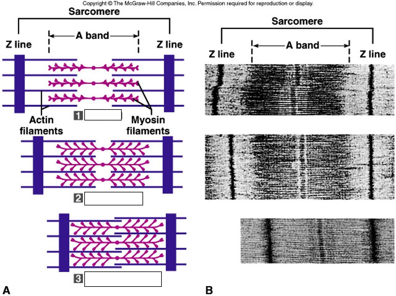
Match the following
spans the length of the thick filament (myosin). Does not change during contraction.
A-Band
spans thick filaments only that shrinks during contraction and lengthens during relaxation.
H-zone
span that includes the thin filaments and z-lines that shortens during contraction and lengths during relaxation.
I-band
“mid-line” of the sarcomere that anchors the thick filaments. During contraction the thin filaments move towards the m-line.
M-Line
the boundary of a sarcomere anchored to the thick filament by titin.
Z-line
Match the following
the end of a nerve cell.
Axon Knob
a neurotransmitter that opens protein channels on the
sarcolemma.
Acetylcholine
(Ach
the process of a vesicle fusing with a cell’s membrane to release chemical substances into the extracellular environment.
Exocytosis
the space between the con terminal bud and the sarcolemma.
Synaptic cleft
an opening of the sarcolemma that allows for the electrical impulse to travel to the sarcoplasmic reticulum
T-Tubules
Reorder the following steps (1-5) of a muscle contraction
Nerve Impulse reaches Neuromuscular Junction
Acetylcholine (ACH) Released by Motor Neuron
ACH binds to Muscle Receptor Sites
Action Potential charge travels down muscle cell
Impulses reaches the SR and Ca+ channels open
Reorder the following (6-10) of the muscle contraction
Ca+ ions diffuse from the SR and bind to troponin molecules
Tropomyosin molecules move and expose specific sites on actin where myosin heads can bind
Cross-bridges form, linking actin and myosin
Actin filaments are pulled toward the center of the sarcomere by pulling of the cross-bridges, increasing the overlap of the thin and thick filaments
The muscle fiber shortens as contractions occurs

Put steps of contraction in order.
rest, nerve action, ACh released, transmitted, Ca released
ACh released, Ca released, nerve action, transmitted, rest
rest, nerve action, Ca released, ACh released, transmitted
ACh released, Ca released, transmitted, nerve action, rest
Where is calcium stored in the muscle cell?
T Tubules
Sarcoplasmic Reticulum (SR)
Sarcolemma
Sarcomere
When the action reaches the muscle cell, it travels down the....
Sarcolemma
T Tubules
Sarcoplasmic Reticulum
Calcium Channel
A calcium deficiency would
create uncoordinated muscle contractions
allow muscle contraction, but not relaxation
prevent contraction by preventing myosin from binding to actin
have no effect on contraction
Another name for the prime mover is
antagonist
agonist
synergist
fixator
A muscle that helps the agonist is called a
antagonist
agonist
synergist
fixator
What is the opposing muscle group for the quadriceps?
biceps
lower back
hamstrings
obliques

In knee flexion, which is the agonist? (the one contracting or getting shorter)
hamstrings
quadriceps
What is the agonist and what is the antagonist in a bicep curl?
biceps are the agonist, triceps are the antagonist
triceps are the agonist, biceps are the antagonist
The muscle that causes the action and does the majority of the work
agonist
antagonist
synergist
fixator
A muscle can be a agonist for one movement and an antagonist for a different movement
true
false
The agonist and antagonist work in __________ to create movement
tandem
random
opposition
synchronization
This enzyme prevents acetylcholine dispersal and activation, therefore, preventing muscle contraction
Calcium
Tropomyosin
Actin
Acetylcholinesterase

Identify the quadriceps
5
8
6
13
Identify the hamstrings
5
8
6
13
Identify the tibialis anterior
5
8
6
13
Identify the gastrocnemius
5
8
6
13
Identify the sternocleidomastoid
1
9
7
10
Identify the trapezius
1
9
7
10
Identify the deltoid
3
2
11
8
Identify the pectorals
3
4
7
12
Identify the biceps
3
4
11
12
Identify the triceps
3
4
11
12
Identify the abdominals
3
4
11
12
Identify the latissimus dorsi
3
4
11
12
This muscle group rotates the head and flexes the neck
Trapezius
Pectorals
Sternocleidomastoid
Latissimus dorsi
This muscle group elevates, depresses, and retracts the scapula
Trapezius
Pectorals
Sternocleidomastoid
Latissimus dorsi
This muscle group abducts the arms
deltoid
biceps
triceps
pectorals
This muscle group flexes the shoulder (arms forward)
deltoid
biceps
triceps
pectorals
This muscle group flexes the elbow
deltoid
biceps
triceps
pectorals
This muscle group extends the elbow
deltoid
biceps
triceps
pectorals
This muscle group bends the torso in a forward direction (think crunches), balance
abdominals
latissimus dorsi
trapezius
sternocleidomastoid
This muscle group adducts the arms
abdominals
latissimus dorsi
trapezius
sternocleidomastoid
This muscle group flexes the hip (legs forward)
quadriceps
hamstrings
tibialis anterior
gastrocnemius
This muscle group extends the hip (legs back)
quadriceps
hamstrings
tibialis anterior
gastrocnemius
This muscle group moves in a dorsiflexion motion
quadriceps
hamstrings
tibialis anterior
gastrocnemius
This muscle group moves in a plantar flexion motion
quadriceps
hamstrings
tibialis anterior
gastrocnemius


What type of contraction is happening here??
isostabilize
isokinetic
isotonic
isometric

Muscle fatigue is...
when the muscle is unable to contract
when you are too tired to exercise
when the muscle is contracted more than 30 secondsd
when O2 comes from direct phosphorylation
What is the correlation between muscle fatigue and oxygen debt?
When O2 decreases, muscle fatigue increases
When O2 increases, muscle fatigue decreases
When O2 is obtained, muscle fatigue decreases
The lesser the oxygen debt, the lesser the muscle fatige
What is the correlation between lactic acid, ATP, and muscle contractions?
increase in lactic acid, decrease ATP, decrease in muscle contraction
increase in lactic acid, decrease ATP, increase in muscle contraction
increase in lactic acid, increase in ATP, decrease in muscle contraction
increase in lactic acid, increase in ATP, increase in muscle contraction
which of the following is true regarding aerobic activity and muscle growth?
increase in mitochondria
increase in vasculature
muscle fiber size DOES NOT increase
Muscle fiber diameter decreases
which of the following is true regarding aerobic activity and muscle growth?
mitochondria number stays the same
decrease in vasculature
muscle fiber size DOES increase
Muscle fiber diameter increases

