
 Worksheets
HBS Mid Year Review (1.1.1-2.1.5)
Total questions: 132
Worksheet time: 2hrs 20mins
Which type of bone cells are responsible for bone formation?
Osteocytes
Osteoblasts
Chondrocytes
Osteoclasts
What is the process called when a broken bone heals and reforms?
bone remodeling
bone regeneration
bone restructuring
bone reconstruction
What is the outer layer of a bone called?
periosteum
epiphysis
medullary cavity
endosteum
What is the scientific term for the collarbone?
humerus
scapula
femur
clavicle
What is the purpose of bone marrow?
To produce and store blood cells
To produce and store fat cells.
To regulate body temperature.
To aid in digestion.
What are the two main types of bone tissue?
dense bone and porous bone
hard bone and soft bone
compact bone and spongy bone
solid bone and hollow bone
What is the function of osteoclasts in bone remodeling?
Bone formation
Bone growth
Bone resorption
Bone repair
A _____ wound might require medical attention whereas a _____ cut or scratch would just need basic bandages.
superficial, deep
deep, superficial
Your lungs are LATERAL to your heart.
True
False
The eyes and nose are on the ______ surface of the body.
posterior
anterior
The stomach is ______ to the heart.
superior
inferior
The hand is PROXIMAL to the shoulder.
True
False
What does it mean when the doctor says he/she is about to dissect the distal end of the popliteal artery?
The doctor is going to dissect an artery at the front of the knee at the end closest to the hip.
The doctor is going to dissect an artery at the front of the knee at the end that is further away from the hip.
The doctor is going to dissect an artery at the back of the knee at the end closer to the hip.
The doctor is going to dissect an artery at the back of the knee at the end that is further away from the hip.
The sternum is ______ to the humerus.
medial
lateral
superior
inferior
Blood, bone, and fat are all examples of _____ tissue.
transport
support
connective
epithelial
If you fracture your ulna, you have broken a bone in your
arm
leg
foot
hand

This pelvis most likely belongs to a
male
female

The bones highlighted in red are called the
tibia
fibula
ulna
femur

Yellow marrow in the bone is mostly
blood plasma
protein
fat
spongy bone

What is angle is being measured by this goniometer?
25
180
155
0

Select the correct range of motion shown in the video clip.
Knee extension
Hip flexion
Knee flexion

Select the correct range of motion shown in the video clip.
Shoulder abduction
Shoulder adduction
Shoulder medial rotation

Select the correct range of motion shown in the video clip.
Shoulder abduction
Shoulder inversion
Shoulder medial rotation

Select the correct range of motion shown in the video clip.
Ankle dorsiflexion
Ankle plantar flexion
Ankle extension
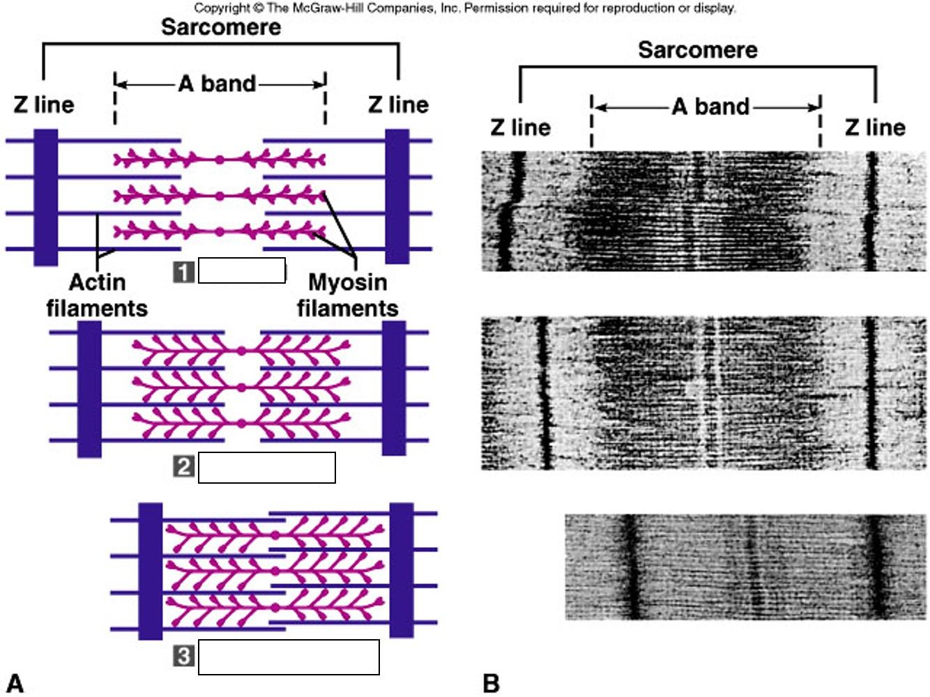
A sarcomere is
a nonfunctional unit of skeletal muscle
a repeating structural unit of skeletal muscle
the area between two intercalated discs
the wavy lines on the muscle seen in the microscope.
Which of the following are composed of myosin?
thick filaments
thin filaments
Z discs
all myofilaments
Which of the following muscle type is not striated?
smooth muscle
skeletal muscle
cardiac muscle
none of above
The bundle of collagen fibers at the end of a skeletal muscle that attaches the muscle to bone is called a(n)
fascicle
tendon
ligament
myofibril
Rigor mortis occurs because
the cells are dead
sodium ions leak out of the muscle
ATP is required to release the attached
actin and myosin molecules
proteins are beginning to break down, thus preventing the flow of calcium ions.
Where is cardiac muscle found?
In the walls of hollow organs
In the walls of the heart
Around the skeleton
In between ribs
What is the function of skeletal muscle?
Movement of the body
Moving food toward the stomach
Pumping blood through the heart
Moving the lungs during breathing

above another part of the body
superior
inferior
distal
proximal
below another part of the body
superior
inferior
distal
proximal
away from point of attachment
superior
inferior
distal
proximal
close to point of attachment
superior
inferior
distal
proximal
back of the body
ventral
dorsal
medial
lateral
toward the surface
superficial
deep
medial
lateral

What is the motion indicated by letter A
Abduction
Extension
Flexion
Hyperextension

What motion is indicated by letter B
Abduction
Extension
Flexion
Hyperextension

What motion is indicated by letter A
PlanterFlexion
Adduction
Abduction
Dorsiflexion

What motion is indicated by letter B
Plantarflextion
Rotation
Inversion
Dorsiflexion

What movement is indicated by Letter A
Rotation
Circumduction
Abduction
Adduction

Select the correct range of motion shown in the video clip.
Ankle dorsiflexion
Ankle plantar flexion
Ankle extension

Select the correct range of motion shown in the video clip.
Shoulder flexion
Shoulder extension
Elbow flexion
Select the correct range of motion shown in the video clip.
Shoulder abduction
Shoulder inversion
Shoulder medial rotation

Select the correct range of motion shown in the video clip.
Hip extension
Hip flexion
Knee flexion

Select the correct range of motion shown in the video clip.
Hip extension
Hip flexion
Knee flexion
Select the correct range of motion shown in the video clip.
Knee extension
Hip flexion
Knee flexion

Select the correct range of motion shown in the video clip.
shoulder extension
shoulder flexion
elbow flexion


What is the surface of the cerebrum called?
cerebral cortex
medulla
axon
What are axons covered in?
myelin sheath
capillaries
What do Axons do?
carry information to the cell body
carry information away from the cell body
What are the components of the central nervous system?
On the cerebral cortex, the grooves or valleys that result from an embryological increase in surface area and folding of the cortex:
sulci
gyri
csf
cord
On the cerebral cortex, the ridges that result from an embryological increase in surface area and folding of the cortex:
hemispheres
sulci
gyri
lobes

These are sections of the brain located behind the frontal lobes; integrate sensory information from the skin, internal organs, muscles, and joints.
Parietal lobes
Frontal lobes
Temporal lobes
Occipital lobes
What are the main parts of the neurons to send and receive information (2 parts)?
nucleus
axon
dendrite
myelin sheath
Node of Ranvier


The Autonomic Nervous System is part of the___________.
Somatic Nervous System
Peripheral Nervous System
Central Nervous System
Sensory Nervous System
The autonomic nervous system is divided into
Motor and Sensory
Peripheral and Central
The Brain and Spinal cord
Sympathetic and Parasympathetic
controls the "fight or flight" response
Sympathetic
Parasympathetic
controls the "rest and digest" response
Sympathetic
Parasympathetic
The PARASYMPATHETIC division of the autonomic nervous system
arouses the body, mobilizing its energy in stressful situations.
calms the body, conserving its energy.
The basic structural and functional unit of brain is
Spinal Cord
Nephron
Neuron
Nerve
Hemisphere that is associated with logical abilities
Hemisphere of the brain that is most associated with artistic abilities
Left
Right
Which of the following structures or systems is correctly paired with its function?
Central nervous system--
processes information and controls responses
Frontal Lobe--portion of the brain that controls body movement
Neurons--
brain cells that control thoughts, calculations, and memory
Peripheral nervous system--releases hormones and controls emotions
Which of the following structures or systems is correctly paired with its function?
Central nervous system--
carries information from the nerves to the muscles and glands
Cerebral cortex--portion of the brain that controls involuntary body movement
Neurons--
brain cells that control thoughts, calculations, and memory
Peripheral nervous system--carries impulses to and from the central nervous system

This portion of the brain is responsible for speech production.
Motor Cortex
Sensory Cortex
Broca's Area
Wernicke's Area

What is the importance of gyri and sulci of the brain?
Decreased surface area
Increase brain size
Decrease neuron connections
Increased surface area

Area of Phineas Gage's brain that was damaged
Frontal lobe
Parietal lobe
Temporal lobe
Occipital lobe
Controls behaviors (hunger, thirst, sleep, body temperature) through the release of hormones.
Hypothalamus
Adrenal Glands
pineal gland
Occipital lobe
Blood Pressure
occipital
Frontal
Brain Stem
Parietal
Part of the brain responsible for vision
Occipital Lobe
Motor Cortex
Temporal Lobe
Pons

The part of the nervous system that consists of the brain and spinal cord
Peripheral Nervous System
Brain Stem
Central Nervous System
Limbic System
Name a disease that involves "tearing" of myelin sheath.

Small cells that engulf and destroy microbes and cellular debris in the CNS
microglia
horns
neuroglia
astrocytes
ATP is required to run
Na/K pumps
leak channels
gated channels
myelin sheaths

Which channels open FIRST at time #2?
leak channels only
gated Na channels
gated K channels
none

Small cells that engulf and destroy microbes and cellular debris in the CNS
microglia
horns
neuroglia
astrocytes
Neuroglial cells that form the fatty myelin sheath on neurons in the CNS are ___________ in the PNS are ________________?
ganglia and oligodendroglia
astrocytes and schwann cells
schwann cells and oligodendroglia
oligodendroglia and schwann cells
Reorder the following (starting at the action potential)
Action potential reached (+30mV)
Na+ inactivation gates close, K+ gates open
K+ exits the cell, repolarization
K+ still open, goes below -70 mV, hyperpolarization
Na/K pump restores to resting potential
Reorder the following (starting at beginning)
Resting potential (-70mV), stimulus received
Na+ gates open, threshold reached (-55 mV)
Na+ gates open
Na+ moves into the axon
Action potential reached (+30mV)
When is the potassium channel open?
during rest only
during repolarization only
during depolarization only
all the time
An electrical impulse within a single neuron is called a
synapse
membrane potential
action potential
cell body
When the membrane potential reaches +30mV, ________ channels open, ________ occurs, and ________ channels close.
sodium, depolarization, potassium
potassium, depoloarization, sodium
sodium, repolarization, potassium
postassium, repolarization, sodium
Which number on the graph represents the threshold potential?
1
2
3
4
6
hyperpolarization
depolarization
repolarization
polarization

At resting membrane potential, neurons have more (a) on the inside of the cell membrane.
At resting membrane potential, neurons have more (a) on the outside of the cell membrane.
Action potentials run along the ______ from ______ to ________.
axon, dendrites, axon terminals
axon, axon terminals, dendrites
dendrites, axon, axon terminals
axon terminals, axon, dendrites

