
 Worksheets
Accessory Structures of the Eye (Anita)
Total questions: 130
Worksheet time: 1hrs 5mins
Which statement best describes the primary function of the eyebrows in ocular protection?
Deflect sweat away from the eyes
Anchor extraocular muscles firmly
Produce basal tears continuously
Focus incoming light onto retina
The palpebrae are responsible for which key role in maintaining ocular health?
Adjusting pupil diameter
Regenerating corneal epithelium
Blinking to spread tear film
Secreting aqueous humor fluid
Which anatomical term identifies the angle where the upper and lower eyelids meet?
Palpebral fissure opening
Lacrimal punctum pore
Palpebral commissure canthus
Conjunctival fornix recess
What structure forms the slit-like opening between the upper and lower eyelids?
Palpebral fissure
Lateral canthus
Conjunctival sac
Medial canthus
Which statement about the conjunctiva is accurate?
Photoreceptor layer detecting images
Transparent mucous membrane lining eyelids
Tough fibrous coat forming sclera
Pigmented ring controlling light entry
Conjunctivitis most directly affects which tissue and typical consequence?
Inflammation of conjunctival membrane
Bacterial infection of lacrimal sac
Redness and irritation of eye surface
Occlusion of lacrimal puncta only
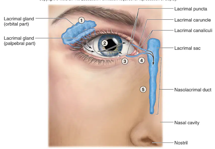
Which structures are part of the lacrimal apparatus involved in tear drainage? Select all that apply.
Lacrimal canaliculi tubes
Lacrimal sac reservoir
Lacrimal puncta openings
Nasolacrimal duct passage
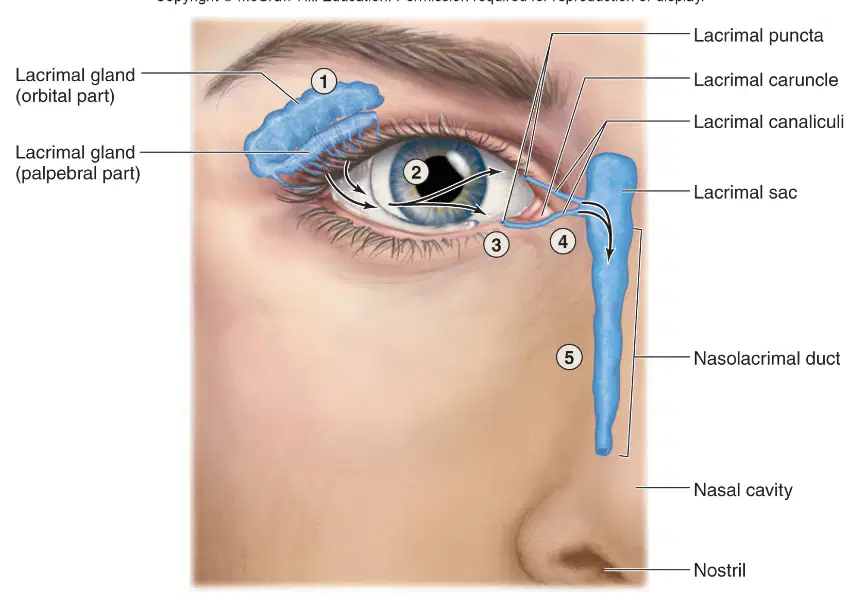
Tears produced by the lacrimal gland typically flow in which correct sequence for drainage?
Canaliculi → puncta → sac → nasal cavity
Puncta → canaliculi → sac → nasolacrimal duct
Sac → canaliculi → puncta → nasolacrimal duct
Nasolacrimal duct → sac → puncta → canaliculi
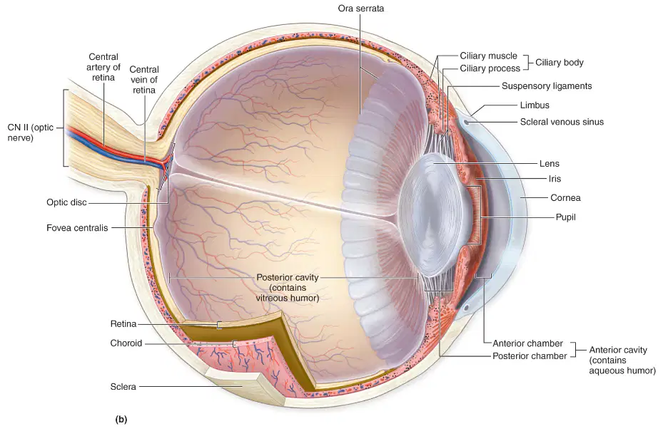
In the external eye diagram, what structure forms the colored ring surrounding the central opening?
Lacrimal caruncle medially
Conjunctiva over cornea
Sclera near the limbus
Iris around the pupil
Which labeled feature is the small pink nodule located at the medial canthus?
Scleral rim
Palpebral fissure
Inferior eyelid
Lacrimal caruncle
Select all external structures that directly contribute to protecting the eye surface from debris and desiccation.
Pupil
Conjunctiva
Superior eyelid
Iris
Inferior eyelid
What term best describes the opening between the superior and inferior eyelids?
Palpebral fissure
Lacrimal canal
Scleral groove
Conjunctival sac
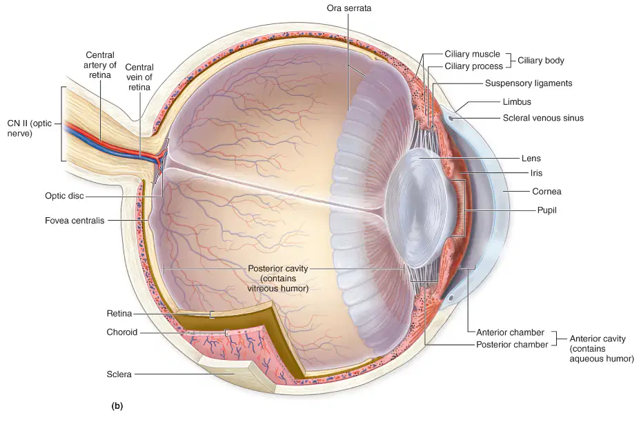
Which structure is the opaque fibrous outer layer visible as the “white of the eye” in the diagram?
Iris pigmented stroma
Conjunctiva vascular film
Pupil central aperture
Sclera forming eye white
Which structure primarily secretes lacrimal fluid to form the aqueous layer of the tear film?
Meibomian glands of eyelids
Goblet cells of conjunctiva
Nasolacrimal duct within maxilla
Lacrimal gland in superolateral orbit
Which components are typical of lacrimal fluid and contribute to antimicrobial defense?
Lysozyme enzyme
Immunoglobulin A
Electrolytes and water
High lipid content
What is a primary function of lysozyme in tears?
Hydrolyze bacterial cell walls
Emulsify lipids in tear film
Stimulate lacrimal gland secretion
Increase oxygen diffusion to cornea
Which combination best describes the roles of the tear film across the cornea?
Pigment deposition and light absorption
Lubrication and optical smoothing
Immune tolerance and vasoconstriction
Muscle attachment and structural support
Arrange the flow of lacrimal fluid from production to drainage into the nasal cavity.
Canaliculi → puncta → lacrimal gland → sac → duct
Lacrimal gland → sac → canaliculi → puncta → duct
Puncta → lacrimal gland → sac → canaliculi → duct
Lacrimal gland → puncta → canaliculi → sac → duct
In the diagram, which structure is primarily responsible for tear production before drainage begins?
Lacrimal sac near medial canthus
Lacrimal gland in superolateral orbit
Nasal cavity receiving tear flow
Nasolacrimal duct within maxilla
Lacrimal puncta on eyelid margin
Which option correctly orders the main pathway of tears as shown in the diagram, from origin to exit?
Lacrimal puncta → lacrimal gland → nasolacrimal duct → nasal cavity
Nasal cavity → nasolacrimal duct → lacrimal sac → puncta
Lacrimal gland → lacrimal puncta → lacrimal sac → nasal cavity
Lacrimal sac → lacrimal gland → nasal cavity → puncta
Select ALL structures that are components of the tear drainage pathway after tears reach the eyelid margin.
Nasolacrimal duct to inferior meatus
Lacrimal sac in lacrimal fossa
Lacrimal puncta at eyelid margin
Lacrimal gland in superolateral orbit
A blockage in which structure would most directly prevent tears from entering the nasal cavity despite normal tear production?
Lacrimal puncta opening medially
Lacrimal sac collecting canaliculi
Lacrimal gland secreting aqueous
Nasal cavity inferior meatus
Nasolacrimal duct descending inferiorly
In the diagram, the lacrimal puncta are positioned to collect tears. Where are they located relative to the eyelids?
Medial eyelid margins
Deep within nasal cavity
Central upper eyelid surface
Lateral canthus region
Posterior to lacrimal sac
Which structure forms the tough outer layer of the eyeball and continues anteriorly as the transparent cornea?
Retina and choroid complex
Sclera continuous with cornea
Iris merging into lens capsule
Ciliary body linked to vitreous
Which combination correctly lists the components of the vascular tunic (uvea)?
Cornea, pupil, vitreous body
Iris, ciliary body, choroid
Sclera, cornea, retina
Lens, optic disc, sclera
The iris performs which primary function related to light entering the eye?
Transduces light into neural signals
Initiates accommodation by thickening lens
Controls pupil diameter via pupillary reflex
Generates aqueous humor in posterior chamber
Which statement accurately differentiates the anterior and posterior chambers of the anterior cavity?
Anterior between sclera and choroid; posterior between lens and retina
Anterior between cornea and lens; posterior between retina and choroid
Anterior between iris and lens; posterior between cornea and iris
Anterior between cornea and iris; posterior between iris and lens
What is the pupil in relation to the iris?
Opening in center connecting two chambers
Photoreceptor layer on iris surface
Muscle ring that forms the lens
Opaque disc behind the iris
Which structures together constitute the fibrous tunic of the eye?
Iris and lens
Cornea and sclera
Ciliary body and choroid
Retina and optic disc
Which statement best describes the optic disc?
Region of maximal cone density
Exit point of optic nerve, no photoreceptors
Ring anchoring suspensory ligaments
Opening that regulates aqueous outflow
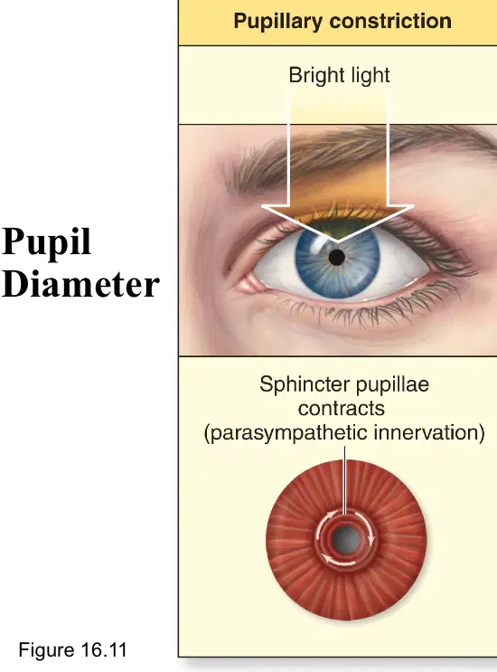
During increased brightness, which response occurs due to the pupillary reflex?
Aqueous humor production increases
Lens flattening to decrease refraction
Pupil constriction reducing light entry
Pupil dilation via radial muscle contraction
Which structures compose the fibrous tunic of the eye?
Choroid and ciliary body
Sclera and iris
Cornea and choroid
Sclera and cornea
Iris and pupil
Primary function of the sclera is to
Protect and maintain globe shape
Absorb excess light internally
Control pupil diameter precisely
Refract most incoming light
Produce aqueous humor continuously
What is the chief role of the cornea?
Provide major light refraction
Anchor extraocular muscles
Supply retinal blood vessels
Secrete vitreous humor
Regulate intraocular pressure
The limbus is best described as the junction between the
Cornea and sclera
Iris and pupil
Choroid and retina
Lens and ciliary body
Vitreous and aqueous
Which regions together constitute the vascular tunic (uvea)?
Iris
Choroid
Sclera
Ciliary body
Cornea
Key function of the choroid includes
Maintaining corneal transparency
Anchoring suspensory ligaments tightly
Producing tears for corneal surface
Generating action potentials in rods
Providing vascular supply to retina
The ciliary body contains which components?
Ciliary muscle
Ciliary processes
Optic disc
Fovea centralis
Suspensory ligaments
Contraction of the ciliary muscle primarily
Moves the optic disc laterally
Flattens the lens for far focus
Dilates the pupil in bright light
Reduces zonular tension for near focus
Increases aqueous outflow at canal
The iris controls the amount of light entering the eye by
Compressing vitreous humor
Thickening the scleral coat
Rotating the lens equator
Adjusting pupil diameter
Shifting the macula lutea
Which statement about pupil control is correct?
Scleral fibers widen the pupil
Sphincter pupillae constricts pupil
Suspensory ligaments dilate pupil
Dilator pupillae constricts pupil
Ciliary processes constrict pupil
Damage at the limbus would most directly affect
Photon capture by photoreceptors
Humor secretion by ciliary processes
Transition between cornea and sclera
Signal exit via optic nerve
Attachment of extraocular muscles
Which pairing is accurate for tunic and primary function?
Vascular—axonal signal conduction
Fibrous—support and protection
Fibrous—phototransduction processing
Vascular—tear film production
Neural—aqueous humor drainage
Increased blood flow to the choroid would most directly enhance
Impulse speed in optic nerve
Aqueous humor formation in angle
Lens accommodation at distance
Light refraction at corneal surface
Nutrient delivery to outer retina
Which structure is NOT part of the vascular tunic?
Uveal tract
Cornea
Iris
Choroid
Ciliary body
A patient with loss of accommodation most likely has dysfunction of the
Rod cells within peripheral retina
Sphincter pupillae within iris
Optic disc fibers exiting globe
Ciliary muscle within ciliary body
Corneal epithelium at limbus
In the pupil diameter regulation diagram, which muscle directly causes constriction of the pupil in bright light?
Ciliary muscle fiber bundles
Sphincter pupillae muscle fibers
Extraocular rectus muscle fibers
Dilator pupillae muscle fibers
Which autonomic pathway primarily drives pupillary dilation when the environment becomes dim?
Enteric neural pathway
Parasympathetic cholinergic pathway
Sympathetic adrenergic pathway
Somatic motor pathway
Select all statements that accurately describe muscle actions during changes in pupil diameter.
Sphincter pupillae contraction reduces aperture size
Sympathetic activity relaxes the sphincter muscle
Dilator pupillae contraction enlarges pupil diameter
Parasympathetic activity contracts the dilator muscle
A patient receives a topical muscarinic agonist in one eye. Which immediate pupillary response is most likely?
Dilation via dilator pupillae contraction
Constriction via sphincter pupillae contraction
No change in diameter from either muscle
Dilation via sphincter pupillae relaxation
Which sequence correctly lists the three principal neural layers of the retina from photoreceptor input to optic nerve output?
Photoreceptors → ganglion cells → bipolar cells
Bipolar cells → photoreceptors → ganglion cells
Photoreceptors → bipolar cells → ganglion cells
Ganglion cells → bipolar cells → photoreceptors
Which structure corresponds to the eye’s blind spot because it lacks photoreceptors?
Macula lutea region with dense cones
Optic disc where axons exit
Fovea centralis within macula
Peripheral retina near ora serrata
Which statements about rods and cones are accurate? Select all that apply.
Cones enable high acuity and color discrimination
Cones dominate the peripheral retina
Rods are concentrated in the fovea centralis
Rods are specialized for scotopic (dim light) vision
Where is visual acuity maximal due to tight packing of cones and minimal convergence?
Optic disc of posterior retina
Fovea centralis of macula lutea
Bipolar cell layer of inner retina
Peripheral retina near ciliary body
Arrange the pathway of signal flow in the retina when light is absorbed by photoreceptors.
Ganglion cells → photoreceptors → bipolar cells
Photoreceptors → bipolar cells → ganglion cells
Bipolar cells → ganglion cells → photoreceptors
Photoreceptors → ganglion cells → optic disc
Which combination best characterizes peripheral retina function?
High rod density, improved motion detection
Equal rod and cone density, uniform acuity
High cone density, sharp central vision
Absence of photoreceptors, blind region
Phototransduction refers to which process in retinal physiology?
Secreting pigment by choroidal cells
Maintaining corneal transparency and oxygen
Converting light into electrical signals
Transporting aqueous humor through the pupil
Which retinal layer’s axons aggregate to form the optic nerve?
Pigmented epithelium
Bipolar cell interneurons
Photoreceptor outer segments
Ganglion cell layer
Which features are typical of rods compared with cones? Select all that apply.
More numerous across retina overall
Poor spatial resolution but high sensitivity
Primarily concentrated within fovea
Incapable of color discrimination
Which description correctly identifies the macula lutea?
Pigmented epithelial layer behind photoreceptors
Central retinal region specialized for detailed vision
Peripheral margin adjacent to ciliary body
Area surrounding optic disc lacking rods
Which statement best distinguishes cones from rods?
Cones mediate color vision and high acuity
Cones are most abundant in peripheral retina
Cones dominate scotopic vision in darkness
Cones project directly to the optic nerve
What is the functional consequence of the optic disc lacking photoreceptors?
Creation of a physiologic blind spot
Enhanced color saturation perception
Improved motion detection peripherally
Increased sensitivity to dim light
Which elements belong to the retina’s neural circuitry between photoreceptors and optic nerve output? Select all that apply.
Ganglion cells generating action potentials
Ciliary processes secreting aqueous humor
Pigmented epithelium absorbing stray light
Bipolar cells relaying graded signals
In the fovea centralis, which property minimizes signal convergence to maximize resolution?
Dense ganglion cell axon myelination
High rod-to-ganglion convergence ratio
Direct cone-to-bipolar-to-ganglion mapping
Absence of bipolar interneurons entirely
Which set correctly matches cell type with primary role in vision?
Ganglion cells: fire action potentials to optic nerve
Bipolar cells: convert light to chemical signals
Photoreceptors: transmit action potentials to brain
Pigment epithelium: relay signals to ganglion cells
Which statement best describes the lens’s role in forming a sharp image on the retina?
It absorbs light to protect photoreceptors
It refracts light to focus on the retina
It converts photons into neural signals
It blocks peripheral rays to reduce glare
During accommodation for near vision, what combination of changes occurs in the eye?
Lens becomes flatter
Lens becomes more rounded
Suspensory ligaments tighten
Ciliary muscle contracts
What happens to suspensory ligaments when the ciliary muscle relaxes?
They slacken and loosen
They detach from the lens
They tighten and pull the lens
They thicken and refract light
Which change most directly increases the refractive power of the lens?
Reducing axial length of the eye
Flattening the lens surfaces
Increasing lens curvature
Dilating the pupil widely
In distant vision, which combination of lens shape and muscle/ligament states is correct in the eye diagram?
Lens flattened; ciliary relaxed; ligaments taut
Lens thickened; ciliary contracted; ligaments taut
Lens flattened; ciliary contracted; ligaments relaxed
Lens thickened; ciliary relaxed; ligaments relaxed
During near vision accommodation, which changes occur simultaneously?
Lens thickens to increase refractive power
Suspensory ligaments become relaxed
Ciliary muscles relax to reduce tension
Lens flattens to reduce refraction
Which statement best explains how ciliary muscle activity alters lens curvature during accommodation?
Relaxation decreases zonular tension, thickening the lens
Contraction increases zonular tension, flattening the lens
Relaxation increases lens curvature by compressing it
Contraction reduces zonular tension, allowing lens to thicken
Which statement best distinguishes vitreous humor from aqueous humor in terms of location?
Vitreous humor fills anterior cavity near cornea
Aqueous humor occupies entire eye except retina
Aqueous humor fills posterior cavity behind lens
Vitreous humor occupies posterior cavity behind lens
Which functions are correctly attributed to aqueous humor? Select all that apply.
Stabilizes retina against detachment
Transmits light to photoreceptors
Maintains intraocular pressure
Nourishes cornea and lens
The vitreous body is primarily composed of which material and serves what key role?
Watery plasma; oxygen delivery to iris
Protein solution; regulates ciliary muscle tone
Gel-like collagen; supports retina structurally
Oil-based fluid; refracts light at cornea
Which structure is directly responsible for producing aqueous humor?
Suspensory ligaments of the lens
Trabecular meshwork and canal of Schlemm
Retinal pigment epithelium cells
Ciliary processes of ciliary body
In the diagram of anterior eye anatomy, where is aqueous humor first secreted before it flows into the anterior chamber?
Trabecular meshwork near sclera
Scleral venous sinus canal
Ciliary processes of ciliary body
Iris surface surrounding the pupil
Which sequence best describes the normal pathway of aqueous humor movement shown in the diagram?
Iris stroma to pupil to scleral venous sinus to posterior chamber
Scleral venous sinus to anterior chamber to pupil to posterior chamber
Ciliary processes to posterior chamber to pupil to anterior chamber
Anterior chamber to pupil to posterior chamber to ciliary processes
Which structures participate primarily in aqueous humor resorption in the diagram? Select all that apply.
Ciliary processes
Pupil opening
Trabecular meshwork
Scleral venous sinus
A blockage at the pupil is most likely to cause which immediate change in aqueous humor dynamics depicted in the diagram?
Impaired flow from posterior to anterior chamber
Increased resorption at scleral venous sinus
Diversion directly into the vitreous chamber
Reduced secretion by ciliary processes
Which statement best describes the outer segment of a vertebrate photoreceptor?
Contains stacked membranous discs with opsins
Houses mitochondria and biosynthetic machinery
Forms synapses onto bipolar and horizontal cells
Pigmented epithelial cells that absorb stray photons
In darkness, what is the dominant ionic current in rod photoreceptors?
Cl− efflux through ligand‑gated channels
Ca2+ efflux via exchanger proteins
K+ influx through voltage‑gated channels
Na+ influx through cGMP‑gated channels
Which changes occur in a rod during phototransduction after photon capture by rhodopsin?
cGMP‑gated channels close
Glutamate release increases
cGMP levels decrease
Membrane hyperpolarizes
Transducin (Gt) is activated
Compared with rods, cones are specialized for which functional characteristics?
High acuity vision
Color discrimination
Fast response kinetics
Greater sensitivity in dim light
Lower threshold for saturation
What is the principal role of the retinal pigmented epithelium adjacent to photoreceptors?
Phagocytoses shed discs and recycles retinal
Generates action potentials for optic nerve
Amplifies light by reflection and scatter
Releases acetylcholine onto ganglion cells
Where are most mitochondria located within a photoreceptor cell?
Synaptic ribbon terminals
Inner segment ellipsoid region
Outer segment disc stack
Pigmented epithelial layer
At the photoreceptor synaptic terminal, decreased glutamate release in light most directly affects which postsynaptic pathway?
Activates ganglion cells via NMDA channels
Inhibits horizontal cells via GABA receptors
Disinhibits ON bipolar cells via mGluR6
Excites OFF bipolar cells via AMPA receptors
In the diagram of visual pathways, which structure carries axons after partial decussation has occurred at the midline?
Optic nerve fibers before crossing
Optic tract fibers after crossing
Lateral geniculate nucleus neurons
Superior colliculus interneurons
Which nuclei are primary relay centers in the thalamus for conscious visual perception?
Optic chiasm commissural neurons
Lateral geniculate nuclei in thalamus
Superior colliculi in tectum
Pretectal nuclei in midbrain
Select all pathways that receive direct retinal input via the optic tract.
Pretectal nucleus
Lateral geniculate nucleus
Primary motor cortex
Superior colliculus
At the optic chiasm, which fibers typically cross to the contralateral side?
Nasal retinal fibers cross contralaterally
Macular fibers always remain ipsilateral
Temporal retinal fibers remain ipsilateral
All retinal fibers cross bilaterally
Which condition results from damage to one optic nerve before the chiasm?
Monocular vision loss in the affected eye
Binocular vision enhancement due to redundancy
Bitemporal hemianopia affecting both temporal fields
Homonymous hemianopia affecting same-side fields
Which structure of the external ear primarily collects and funnels sound waves toward the ear canal?
Tympanic membrane at canal end
Auricle projecting from head
Ceruminous glands in skin
Stapes within middle ear
What is the main role of the external acoustic meatus in hearing?
Equalizes middle ear pressure
Transduces signals to CN VIII
Channels sound to eardrum
Amplifies ossicle vibrations
Cerumen is produced by ceruminous glands. Which function best describes cerumen?
Detects frequency differences
Moves stapes at oval window
Traps debris, protects canal
Lubricates ossicular joints
Which statement about the tympanic membrane is accurate?
It secretes protective wax
It is part of the cochlear duct
It houses hair cell stereocilia
It vibrates with sound waves
Which cranial nerve carries auditory information from the cochlea to the brainstem?
Vestibulocochlear CN VIII
Trigeminal nerve CN V
Glossopharyngeal CN IX
Facial nerve CN VII
Select all components that belong to the external ear.
External acoustic meatus
Malleus
Cochlear duct
Auricle
Tympanic membrane
Which event immediately follows vibration of the tympanic membrane?
Deflection of stereocilia
Movement of auditory ossicles
Activation of vestibular hair cells
Opening of round window
Which pairing correctly matches structure with role in the sound pathway shown?
Auricle—transduces to neural code
Eardrum—initiates ossicle motion
Cerumen—amplifies wave pressure
CN VIII—equalizes ear pressures
Which structure connects the middle ear to the nasopharynx to equalize pressure across the tympanic membrane?
Oval window of the cochlea
External acoustic meatus
Auditory tube (Eustachian tube)
Round window of the cochlea
In the chain of auditory ossicles, which bone directly contacts the oval window to transmit vibrations into the inner ear fluid?
Stapes footplate in oval window
Stapedius tendon on stapes
Incus linking malleus and stapes
Malleus attached to tympanic membrane
Which statements correctly describe the roles of the oval and round windows during sound transmission?
Oval window receives stapes vibrations
Round window dissipates pressure waves
Oval window equalizes middle ear pressure
Round window initiates ossicle movement
What is the primary function of the tympanic cavity as it relates to hearing?
Pressure reservoir in semicircular ducts
Sensory epithelium with hair cells
Fluid-filled cochlear duct
Air-filled chamber housing ossicles
Select all correct statements about the auditory ossicles.
Malleus contacts the tympanic membrane
Incus forms a lever between malleus and stapes
Ossicles equalize pressure via the round window
Stapes transmits force to the oval window
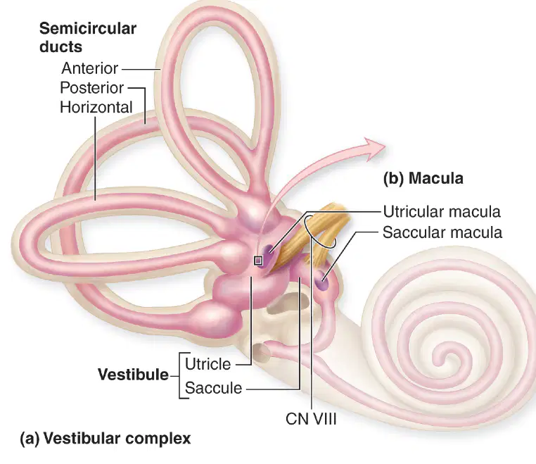
Which inner ear region contains the utricle and saccule housing maculae for sensing static equilibrium and linear acceleration?
Cochlea spiral labyrinth
Membranous cochlear duct
Vestibule central chamber
Anterior semicircular canal
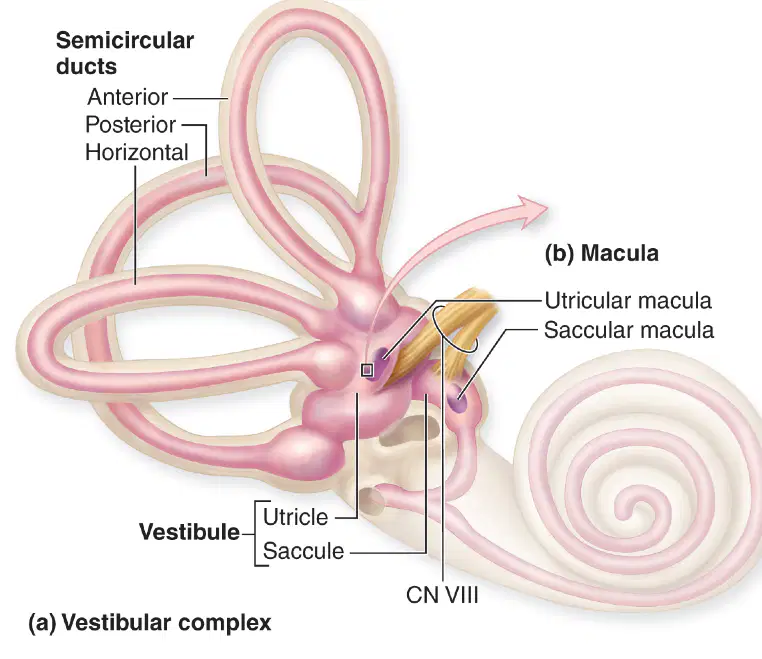
Select all structures that are part of the vestibular apparatus responsible primarily for angular acceleration detection.
Anterior semicircular duct
Posterior semicircular duct
Horizontal semicircular duct
Membranous cochlear duct
Which pairing correctly matches an inner ear region with a principal function?
Cochlea — detect sound via hair cells
Membranous cochlear duct — measure angular acceleration
Vestibule — amplify tympanic vibrations
Semicircular canals — transmit signals through CN VII
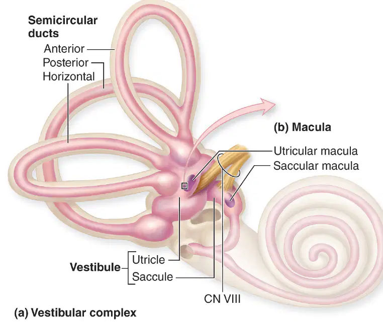
In the membranous labyrinth, which ducts lie within the semicircular canals and contribute to detecting head rotation?
Saccular macula plates
Utricular macula plates
Membranous semicircular ducts
Membranous cochlear duct
Which fluid fills the membranous labyrinth within the cochlea and bathes hair cell stereocilia?
Perilymph in scala tympani
Interstitial fluid of modiolus
Endolymph within cochlear duct
Perilymph within vestibule
In the bony labyrinth of the inner ear, which two spaces specifically contain perilymph around the cochlear duct?
Scala tympani
Scala media
Scala vestibuli
Vestibular aqueduct
Which structure houses the sensory epithelium that transduces sound within the cochlear duct?
Spiral organ on basilar membrane
Macula within utricle wall
Crista in semicircular canal
Tectorial membrane alone
The otolithic membrane shown in the macula diagram is embedded with which inert particles that add mass?
Otoliths composed of calcium carbonate
Endolymph lipid droplets
Kinocilium crystals
Tectorial proteoglycan rods
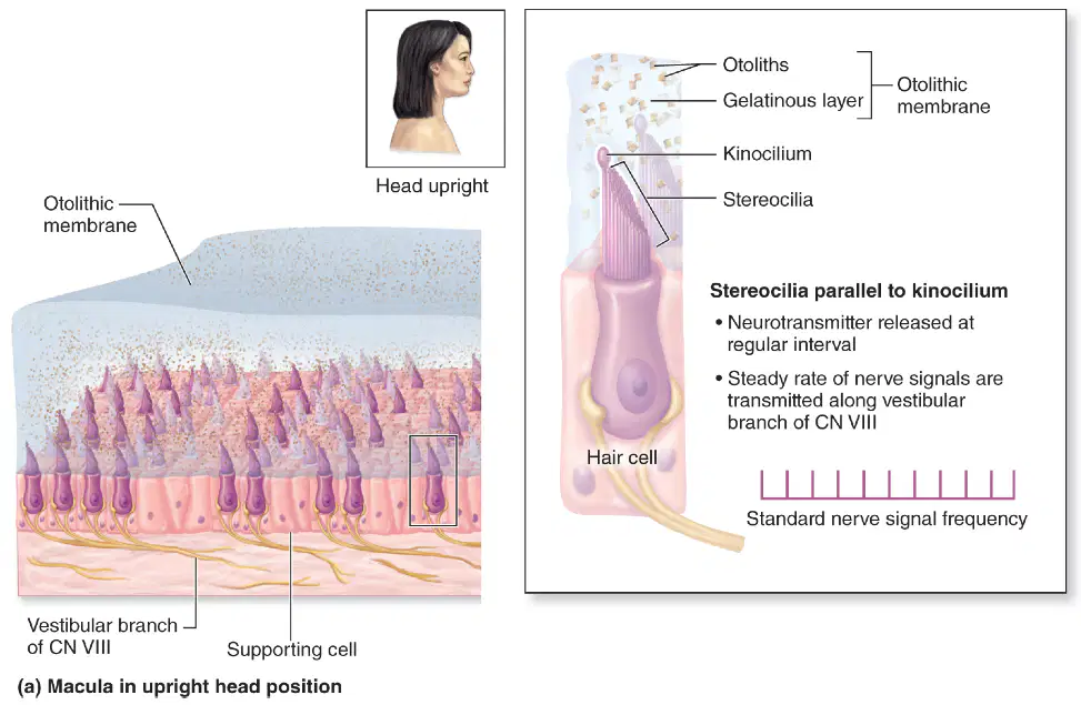
On a vestibular hair cell of the macula, stereocilia oriented parallel to the kinocilium at rest lead to which neural output?
Maximal excitation bursts only
Steady baseline firing of CN VIII
No transmitter release occurs
Complete inhibition of vestibular nerve
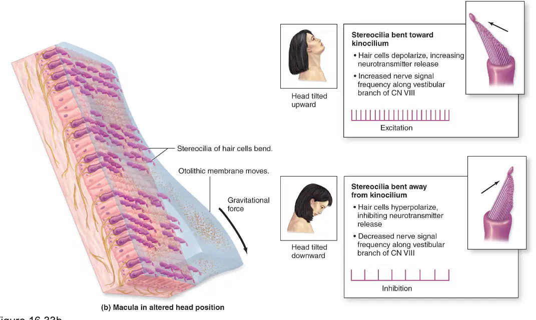
When the head tilts upward, the otolithic membrane shifts such that stereocilia bend toward the kinocilium. What is the immediate hair cell response?
Hyperpolarization with decreased firing
Depolarization increasing neurotransmitter release
No change due to adaptation
Mechanical detachment of kinocilium
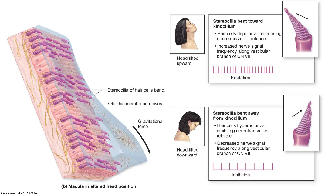
Head tilt downward bends stereocilia away from the kinocilium. Which change occurs in vestibular nerve signaling?
Signal frequency remains unchanged
Phase-locked firing to sound waves
Signal frequency increases markedly
Signal frequency decreases below baseline
Which pair correctly matches labyrinth component with its contained fluid?
Membranous labyrinth—endolymph
Cochlear duct—perilymph
Bony labyrinth—endolymph
Membranous labyrinth—perilymph
Within the cochlea, where is the spiral organ situated relative to surrounding scalas?
On Reissner's membrane of scala vestibuli
On tectorial membrane of scala tympani
On basilar membrane of scala media
Within osseous spiral lamina only
Which statement best distinguishes the functions of vestibule and semicircular canals?
Vestibule detects linear acceleration and position
Semicircular canals detect linear acceleration
Vestibule detects angular acceleration
Both detect only auditory frequencies
In the macula diagram, which cells provide structural support adjacent to hair cells?
Supporting cells in the epithelium
Type I afferent neurons
Schwann cells of cochlear nerve
Osteocytes of bony labyrinth
During excitation in macular hair cells, which ion movement most directly produces depolarization?
Sodium influx from perilymph
Potassium influx from endolymph
Chloride efflux through stereocilia
Calcium efflux to tectorial membrane
Which structure first collects and funnels sound waves toward the external acoustic meatus in the diagram?
Auricle of the outer ear
Tympanic membrane surface
Oval window of cochlea
Basilar membrane region
In the depicted pathway, what is the immediate consequence of tympanic membrane vibration?
Auditory ossicles amplify motion
Basilar membrane displaces upward
Hair cells release neurotransmitter
Cochlear duct receives endolymph
Which events together transduce mechanical energy into neural signals at the end of the pathway?
Basilar membrane displacement
Auricle funneling of sound
Hair cells stereocilia distortion
Pressure waves at the oval window
Where do pressure waves enter the cochlea after ossicular amplification according to the diagram?
Round window into scala tympani
Oval window into scala vestibuli
Eustachian tube into vestibule
External meatus into cochlear duct
Which sequence best represents the mechanical-to-neural pathway shown?
Auricle → meatus → tympanic membrane → ossicles → oval window → scala vestibuli → cochlear duct → basilar membrane → hair cells → cochlear branch CN VIII
Auricle → meatus → ossicles → tympanic membrane → round window → scala tympani → cochlear duct → kinocilium → vestibular branch CN VIII
Auricle → tympanic membrane → ossicles → oval window → vestibule → semicircular canals → cupula → hair cells → vestibular branch CN VIII
Meatus → auricle → tympanic membrane → ossicles → oval window → scala tympani → utricle → basilar membrane → hair cells → facial nerve
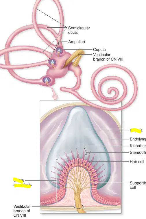
Which structures detect angular acceleration of the head during rotation?
Utricle and saccule maculae
Semicircular duct ampullae
Vestibular branch ganglia
Cochlear spiral organ
Static equilibrium and linear acceleration are primarily sensed by which vestibular components?
Crista ampullaris in ampullae
Olfactory epithelium in nasal cavity
Maculae of utricle and saccule
Organ of Corti in cochlea
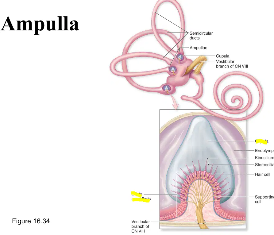
In the ampulla, bending of stereocilia toward the kinocilium causes what immediate effect on hair cells?
Hyperpolarization decreases neurotransmitter release
No change because endolymph is stationary
Depolarization increases neurotransmitter release
Mechanical inhibition of cupula movement
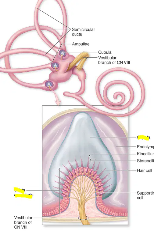
Which set correctly matches structure to function in the vestibular apparatus?
Utricle—detects vertical linear acceleration
Cupula—gel structure displaced by endolymph flow
Ampulla—houses crista ampullaris for angular acceleration
Saccule—detects horizontal linear acceleration

