
 Worksheets
Skull & Sinus positioning
Total questions: 112
Worksheet time: 59mins

In which of these radiographs is the CR directed 15° caudal and exiting at the nasion?
A
B
C
D

In which of these radiographs will the CR be directed 30° caudal when the patient's OML is perpendicular with the plane of the IR?
A
B
C
D

In which of these radiographs will the CR be directed perpendicular 2" superior to the EAM?
A
B
C
D

In which of these radiographs are the petrous ridges completely filling the orbits.
A
B
C
D

In which of these radiographs is the dorsum sellae & posterior clinoid process demonstrated within the foramen magnum?
A
B
C
D
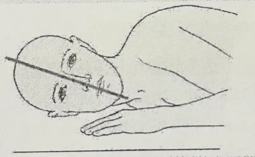
Describe what type of patient is represented in the diagram, then explain how the position of the patient should be adjusted to have the MSP aligned properly with the IR.
Hyposthentic/Asthenic
Hypersthenic
Place cushion under chest area
Place cushion under head
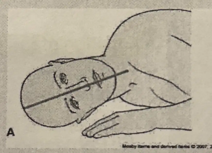
Describe what type of patient is represented in the diagram, then explain how the position of the patient should be adjusted to have the MSP aligned properly with the IR.
Hyposthenic/Asthenic
Hypersthenic
Place cushion under chest area
Place cushion under head

Which of the diagrams shown demonstrates a skull that is short from front to back, wide from side-to-side and shallow from vertex to base (brachycephalic)?
A
B
C

Which of the diagrams shown correctly demonstrates a patient with a dolichocephalic skull?
A
B
C

In order to correctly place the patient's OML perpendicular with the plane of the IR on a hypersthenic patient, which of the following should be implemented?
A
B
C
Which plane of the head is placed PARALLEL to the plane of the IR for a lateral projection of the skull?
Sagittal
Transverse
Midsagittal
Midcoronal
The CR and center of the IR position for a LATERAL projection of the skull is:
1 inch below the EAM
2 inches below the EAM
1 inch above the EAM
2 inches above the EAM
Which of the following is true regarding the lateral projection of the skull?
1.)the midsagittal plane of the head is parallel to the plane of the IR
2.)the interpupillary line is perpendicular to the IR
3) the mentomeatal line is parallel with the bottom edge of the IR
1 & 2
1 & 3
2 & 3
1,2, and 3
Which of the following should be seen superimposed on a lateral projection of the skull?
1.)orbital roofs
2) external acoustic meati
3.) temporomandibular joints
1 & 2
1 & 3
2 & 3
1,2, and 3
Which method of examining the skull will demonstrate the petrous ridges in the orbits, the ethmoid and frontal sinuses, and the crista galli?
Towne
Caldwell
Schuller
Rhese
The CR angle for the PA Axial (Caldwell) projection of the skull is:
5 degrees cephalad
10 degrees cephalad
12 degrees caudad
15 degrees caudad
Which of the following is perpendicular to the plane of the IR for a Caldwell projection of the skull?
Glabellomeatal line
Acanthoeatal line
Orbitomeatal line
Mentomeatal line
Often a patient cannot be turned into the prone position for a PA axial projection of the skull (Caldwell method). What CR angle could be used if the AP axial projection is used instead?
10 degrees caudad
15 degrees cephalad
10-15 degrees caudad
10-15 degrees cephalad
Which of the following lines is placed perpendicular to the plane of the IR for the AP axial (Towne) projection?
Orbitomeatal line
Infraorbitomeatal line
Glabellomeatal line
Acanthiomeatal line
If the patient cannot flex the neck to place the orbitomeatal line perpendicular to the IR for a Towne projection, which line should be placed perpendicular?
Acanthiomeatal line
Infraorbiomeatal line
Glabellomeatal line
Mentomeatal line
Which of the following is true regarding the placement of the IR for a Towne projection of the skull?
1.) its upper margin is at the level of the top of the cranium
2.)its upper margin is 2 inches above the top of the cranium
3.)its upper margin is 2 inches below the top of the cranium
1 only
2 only
3 only
1,2, and 3

Haas
Towne
Rheese
Caldwell
If the infraorbitometal line is placed perpendicular to the IR during an AP axial (Towne) projection of the skull, how much is the CR angled?
15 degrees caudad
30 degrees caudad
37 degrees caudad
45 degrees caudad
Which of the following is clearly demonstrated within the foramen magnum during an AP axial (Towne) projection of the skull?
1.)dorsum sellae
2) sella turcica
3.) posterior clinoid processes
1 & 2
1 & 3
2 & 3
1,2, and 3
What is the CR angulation for demonstrationed of the entire foramen magnum during a Towne projection?
37 degrees caudad
40 degrees caudad
60 degrees caudad
40-60 degrees caudad
Which of the following methods will clearly demonstrate the petrous ridges, foramen magnum, dorsum sellae, and posterior clinical processes?
1)Haas (PA axial)
2) Towne (AP axial)
3) Schuller (SMV)
1 & 2
1 & 3
2 & 3
1,2, and 3
What is the average CR angulation for the PA axial (Haas) projection of the skull?
25 degrees caudad
25 degrees cephalad
30 degrees caudad
30 degrees cephalad
Which line should be placed parallel to the plane of the IR for the SMV projection of the cranial base?
Acanthiomeatal line
Orbitomeatal line
Infraorbitomeatal line
Mentomeatal line
Radiographic demonstration of the cranial base is performed by which method?
Haas
Rhese
Towne
Schuller
What is the CR angulation for the SMV projection?
0 degrees
5 degrees caudad
5 degrees cephalad
5-7 degrees cephalad
For an SMV projection of the cranial base, the CR should always be perpendicular to the:
Mentomeatal line
Orbitomeatal line
Infrorbitomeatal line
Acanthiomeatal line
Which of the following are clearly demonstrated on an SMV projection of the cranial base?
1)mastoid process
2)sphenoid sinuses
3)carotid canals
1 & 2
1 & 3
2 & 3
1,2, and 3
What is the CR entrance point for the lateral projection of the sella turcica?
At the EAM
2 inches above the EAM
2 inches below the EAM
3/4 inch anterior to and superior to the EAM

SMV
VSM
AP Axial
PA Axial
Patients who are having an examination of the paranasal sinuses should always be examined in the upright position to:
1) demonstrate the presence or absence of fluid
2) differentiate between fluid and other pathological conditions
3) make it easier for the radiographer to position the patient
1 & 2
1 & 3
2 & 3
1,2, and 3
For a LATERAL projection of the parental sinuses the CR is directed:
1 inch posterior to the outer canthus
2 inch posterior to the outer canthus
1 inch anterior to EAM
2 inches anterior to EAM
Which of the following should be clearly demonstrated on a lateral projection of the paranasal sinus?
1)all four sinus groups
2)superimposed orbital roofs
3)superimposed mandibular rami
1 & 2
1 & 3
2 & 3
1,2, and 3
Which parts of the patient's face touch the table for a PA axial projection (Caldwell method)?
1)forehead
2)nose
3) chin
1 & 2
1 & 3
2 & 3
1,2, and 3
Which of the following are demonstrated on an AP axial (Towne method) projection of the skull?
1) petrous pyramids
2) occipital bone
3) posterior portion of the foramen magnum
1 & 2
1 & 3
2 & 3
1,2, and 3
In which projection of the sinuses will all four sets of sinuses be demonstrated?
Lateral
Pa
Pirie
Waters
In which projection of the sinuses will the petrous pyramids be projected below the floor of the maxillary sinuses?
Waters/ Parietoacanthial
Lateral
PA Caldwell
Pirie
Which sinuses are best demonstrated on the Pirie Method?
Sphenoid
Ethmoid
Maxillary
Frontal
Where does the CR exit for the Waters projection?
Glabella
Nasion
3” superior to nasion
Acanthion
For the Waters method, if the patient's chin is overextended/ underextended the petrosa are projected over the inferior portions of the maxillary sinuses and can obscure underlying pathology.
Overextended
Underextended
Name the projection and the SID which should be employed for preoperative measurement of the sinuses?
Lateral
72” SID
40” SID
Pirie
List the routine projections for the paranasal sinuses routine:
PA Caldwell
Lateral of affected side
Waters
Pirie
PA

Which of the diagrams will best demonstrate the frontal and anterior ethmoid sinuses?
A
B
C
D

Which of the diagrams will best demonstrate all four sets of sinuses?
A
B
C
D

Which of the diagrams will best demonstrate the petrous ridges below the maxillary sinuses?
A
B
C
D

Which of these diagrams should be performed to demonstrate the ethmoid and sphenoid sinuses?
A
B
C
D

In which of these diagrams does the OML form a 37 degree angle with the plane of the IR?
A
B
C
D

Which of these diagrams is not part of our routine for the sinus series?
A
B
C
D

In which of these diagrams should the CR exit at the acanthion?
A
B
C
D

Which of the radiographs shown above is underexposed?
A
B
C

Which of the following Radiographs shown above is correctly exposed?
A
B
C

Which of the radiographs shown above is overexposed?
A
B
C

A
B
When positioning for the parietoacanthial projection of the sinuses, if the maxillary sinuses are foreshortened and the antral floors are not demonstrated, the patient's chin was underextended/ overextended.
Underextended
Overextended
True or False
For the PA Axial transoral projection, the CR should be angled 15° caudal and exit through the open mouth, to best demonstrate the sphenoid sinuses projected in the middle of the open mouth.
True
False
If the patient were unable to stand which of the following positions could be employed in order to demonstrate air/fluid levels in the sinuses?
Lateral recumbent position
Dorsal recumbent position
Dorsal decubitus position
Ventral recumbent position

Name the projection
Waters method
Right Lateral
Pirie

Name the projection:
Right lateral sinuses
Waters method
Left lateral sinuses
List the routine for a skull series
PA
PA Caldwell
Townes
Right Lateral
Left Lateral
For the SMV projection, the OML/IOML SHOULD BE Parallel/Perpendicular to the film?
OML
IOML
Parallel
Perpendicular
List the routine for a partial skull series
Townes
PA Caldwell
Lateral of affected side
PA
AP
List the routine for the Sella Turcica series
Haas (or Townes)
Lateral
PA
PA Caldwell
The degree of difference between the OML and IOML is:
7 degrees
8 degrees
The degree of difference between the OML and GML is
7 degrees
8 degrees
What should the patient be instructed to do in order to place the OML perpendicular with the plane of the IR, when the patient is supine?
Tuck your chin
Bend your head back

& what is the projection called?
A
B
C
PA
PA AXIAL (Caldwell)

& what is the projection called?
A
B
C
PA AXIAL (Caldwell)
PA

& name the projection
A
B
C
Lateral Dorsal Decubitus
RAO

& what is the name of the projection?
A
B
C
AP Reverse Caldwell
PA

1.)
Townes
SMV
Caldwell
Haas

2.)
SMV
Townes
Caldwell
Haas

3.)
Caldwell
Townes
SMV
Haas

4.)
Haas
Townes
SMV
Caldwell

Which of these positioning diagrams is not included in the routine for Skull?
A
B
C
D

In which of these positioning diagrams should the patient's IPL be perpendicular with the plane of the IR?
A
B
C
D

In which of these positioning diagrams is the patient's IOML placed parallel with the plane of the IR and perpendicular with the CR?
A
B
C
D

In which of these diagrams would the central ray be angled 37° if the OML cannot be perpendicular with the plane of the IR?
A
B
C
D

In which of these diagrams is the patient's MSP parallel w/ the IR, IPL perpendicular w/ thejR & the IOML parallel w/the bottom of the IR?
A
B
C
D
E

In which of these diagrams is the CR parallel to the patient's OML?
A
B
C
D
E

Which of the diagrams best demonstrates the frontal bone as well as the anterior ethmoid air cells?
A
B
C
D
E

In which of these diagrams will the petrous ridges be projected in the orbits?
A
B
C
D
E

In which of these diagrams will the petrous ridges be projected in the lower 1/3 or the orbits?
A
B
C
D
E

In which of the diagrams will the mandibular condyles be placed anterior to the petrous ridges?
A
B
C
D
E
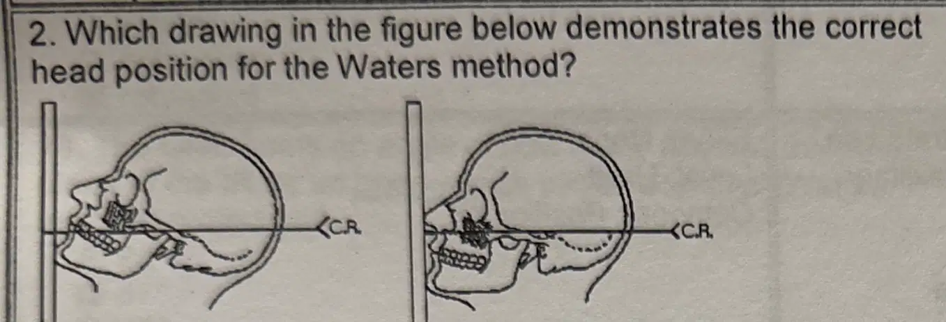
Left
Right
For the open-mouth modification of the Waters method, the CR should exit the:
Nasion
glabella
Acanthion
Open Mouth
To successfully demonstrate the ethmoidal and sphenoidal sinuses on the SMV projection, which of the following must occur?
1) the patient must be recumbent
2) the infraorbitomeatal line must be parallel with the IR
3)the CR must be perpendicular to the infraorbitomeatal line and horizontal
1 & 2
1 & 3
2 & 3
1,2, and 3
At which level should the CR enter the base of the skull for the SMV projection of the sinuses?
3/4 inch posterior to the level of the EAM
3/4 inch anterior to the level of the EAM
3/4 inch below the mental protuberance
1 inch below the mental protuberance
Which projections will demonstrate the ethmoidal sinuses?
1.)PA
2) PA axial (Caldwell)
3) SMV
1 & 2
2 & 3
1 & 3
1,2, and 3
The PA projections of the sinuses can demonstrate which sinuses?
1) ethmoidal
2) sphenoidal
3) maxillary
1 & 2
1 & 3
2 & 3
1,2, and 3
Which line is placed perpendicular to the plane of the IR for the PA projection of the sinuses?
Glabellomeatal
Orbitomeatal
Acanthiomeatal
Infraorbitomeatal
The respiration phase for all projections of the facial bones and sinuses is:
Suspended
Inspiration
Expiration
Shallow breathing
To demonstrate the sphenoidal sinuses on the PA projection, the CR should be angled:
10 degrees caudad
10 degrees cephalad
15 degrees caudad
15 degrees cephalad
Which of the following should be clearly demonstrated on the PA projection for specific demonstration of the maxillary sinuses?
1)maxillary sinuses below the petrous ridges
2) sphenoidal sinuses visible through the frontal bone
3.) close beam restriction of the sinus area
1 & 2
1 & 3
2 & 3
1,2, and 3
Which reference line is positioned horizontal to ensure proper extension of the head during a lateral projection of the sinuses?
AML
OML
IOML
MML
Which reference line is positioned perpendicular to the angled IR for the PA axial (Caldwell method) projection of the sinuses?
AML
OML
MML
IOML
The central ray forms an angle of how many degrees with the OML for the PA axial (Caldwell) projection of the sinuses?
5 degrees
12 degrees
15 degrees
20 degrees
Which of the following reference lines is placed perpendicular to the IR for a parietoacanthial (Waters method) projection?
MML
OML
AML
IOML
Where is the IR centered for the parietoacanthial (Waters method) projection of the sinuses?
Nasion
Inion
Glabella
Acanthion
Where are the petrous ridges seen on a parietoacanthial (Waters method) radiograph?
I’m the middle of the maxillary sinuses
Superior to the maxillary sinuses
Inferior to the floor of the maxillary sinuses
In the lower 2/3rds of the maxillary sinuses
The OML forms an angle of how many degrees from the plane of the IR for an open-mouth parietoacanthial (Waters method) projection?
25 degrees
37 degrees
45 degrees
Which of the following foramina will be demonstrated in the orbit on a parietoacanthial (Waters method) radiograph?
Ovale
Jugular
Spinosum
Rotundum
The CR is directed perpendicular to which reference line for the SMV projection of the sinuses?
OML
AML
MML
OIML
Which projection will best demonstrate the frontal and anterior ethmoidal sinuses?
1) PA
2) parietoacanthial (Waters)
3) PA axial (Caldwell)
1 only
2 only
3 only
1,2, and 3
When using the angled grid technique for the PA axial (Caldwell method) projection of the sinuses, the vertical grid device must be angled:
7 degrees
10 degrees
15 degrees
20 degrees
Which of the following must be projected below the maxillary sinuses for the parietoacanthial projection (Waters method) of the sinuses?
Zygomatic bones
Petrous pyramids
Inferior orbital margin
Sphenoidal sinuses
The parietoacanthial projection (Waters method) of the sinuses requires the orbitomeatal line to be placed how many degrees from the plane of the IR?
20 degrees
27 degrees
30 degrees
37 degrees
Which sinus is projected through the mouth on the open-mouth modification of the Waters method?
Frontal
Ethmoidal
Sphenoidal
Maxillary
Which of the following projections will best demonstrate the maxillary sinuses?
1) parietoacanthial (Waters)
2) PA axial (Caldwell)
3) submentovertical
1 only
2 only
3 only
1,2, and 3

