
 Worksheets
HAP 1st Semester Review
Total questions: 200
Worksheet time: 2hrs 17mins
toward the midline of the body
distal
lateral
medial
inferior
toward or at the front of the body
anterior
dorsal
deep
distal
at the body surface
distal
superficial
deep
proximal

Toward the head end of upper part of body
Medial
Inferior
Lateral
Superior

Away from the midline
Medial
Lateral
Distal
Proximal

Toward a structure's origin or point of attachment to the trunk
Lateral
Proximal
Distal
Medial
Away from the head.
Inferior
Lateral
Deep
Distal
Toward the back of the body.
Superficial
Distal
Superior
Posterior
Away from the trunk.
Superior
Distal
Lateral
Anterior
Away from the surface of the body.
Medial
Inferior
Proximal
Deep
back of the body
ventral
dorsal
medial
lateral
Posterior
behind
in front
away from midline
further from trunk
Distal
toward the body surace
further from the trunk
away from the body surface
below
Superior
below
above
in front
behind
Front of the body
Ventral/ Anterior
Dorsal/ Posterior
Superior
Sagittal
The thigh is ____ to the shoulder.
distal
inferior
superior
posterior
The brain is ____ to the skull.
proximal
superficial
deep
lateral

Which letter indicates lateral?
F
E
A
G
Which letter indicates proximal?
H
G
C
E
Which letter indicates anterior?
H
G
C
F
Front of knee
popiteal
plantar
patellar
cervical
brachial
arm
forearm
wrist
hip
buccal
wrist
cheek area
curve of shoulder
thigh
cervical
neck region
hip
anterior of leg
curve of shoulder
coxal
the shin
hip
neck region
thigh
digital
point of shoulder
fingers and toes
anterior of leg
cheek area
femoral
upper arm area
thigh
lower anterior of leg
eye area
frontal
forehead
eye area
chin
mouth
inguinal
chin
lateral part of leg
forehead
area where thigh meets body trunk
orbital
genital region
eye area
forehead
chin
pectoral
anterior knee
occuring on the chest
ribs
arm
occipital
posterior of head
below the elbow
eye area
between shoulder blades
fibular
lateral part of leg
front of head
lower area of arm
eye area
lumbar
lower back area
lower leg area
posterior of leg
shoulder blade
vertebral
spine
lower leg
neck
behind the head
The study of the structure and shape of the body and its parts and their relationships to one another is PHYSIOLOGY.
True
False
The study of how the body and its parts work or function is PHYSIOLOGY
True
False
The study of how the body and its parts work or function is PHYSIOLOGY
True
False
cells→ ___→ organs→ organ systems→ organism
What can fit in the blank?
Macromolecules
Cell Products
Proteins
Tissues
The highest level of structural organization.
Cell
Organ System
Organism
This system is the external covering of the body.
Endocrine
Integumentary
Lymphatic
Stimulus, Efferent Pathway, Response, Input, Output, Afferent Pathway, Receptor, Control Center
What is the correct order?
Receptor, Stimulus, Input, Efferent Pathway, Control Center, Output, Afferent Pathway, Response
Stimulus, Receptor, Input, Efferent Pathway, Control Center, Output, Afferent Pathway, Response
Stimulus, Receptor, Input, Afferent Pathway, Control Center, Output, Efferent Pathway, Response
Receptor, Stimulus, Input, Afferent Pathway, Control Center, Output, Efferent Pathway, Response
The heart is located ____ to the diaphragm.
Superior
Inferior
Medial
Lateral
The muscles are ____ to the skin.
Superficial
Proximal
Distal
Deep
The shoulder JOINT is ____ to the elbow JOINT.
Proximal
Distal
Superior
Inferior
In ANATOMICAL POSITION, the thumb is ____ to the index finger.
Medial
Lateral
Superior
Inferior
The vertebral region is _______ to the scapular region.
Lateral
Medial
Superior
Inferior
Anatomical terms that apply to the backside of the body when it is in anatomical position:
Ventral and Anterior
Back and Rear
Posterior and Dorsal
Cephalic and Lateral

Is this an example of negative or positive feedback?
Negative
Positive
A sagittal section cuts the body into ____ and ____ parts.
Right and Left
Superior and Inferior
Anterior and Posterior
A frontal section divides the body into ____ and ____ parts.
Right and Left
Superior and Inferior
Anterior and Posterior
A transverse or cross section divides the body into _____ and ____ parts.
Right and Left
Superior and Inferior
Anterior and Posterior
The topics of human anatomy and physiology are difficult to separate because the structures of the body parts are closely related to their
chemicals
function
forces
water
Homeostasis refers to
changing external
conditions.
stable external
conditions.
changing internal
conditions.
maintaining
internal conditions.
all of the
above.
Which of the following examples illustrates a homeostatic mechanism?
shivering in
response to a drop in body temperature
increasing body
temperature during exercise
decreasing body
temperature during prolonged exposure to cold conditions
dehydration from
lack of water intake
frostbite on exposure to cold
The diaphragm separates the thoracic and the abdominopelvic cavities.
True
False
The mediastinum separates
the thoracic
cavity from the abdominal cavity.
the thoracic
cavity into right and left parts.
the thoracic
cavity from the pelvic cavity.
the abdominal
cavity from the pelvic cavity.
the abdominal
cavity into right and left parts.
Which of the following organs is found in the pelvic cavity?
urinary bladder
Kidneys
liver
spleen
gallbladder
The membrane on the surface of the lung is called the
visceral
pleura.
parietal
pleura.
visceral
pericardium.
parietal
pericardium.
The elbow is distal to the wrist.
True
False
The stomach is an example of which organizational level?
molecule
organelle
tissue
organ
Deepest layer (not actually part of the integument) that is made of elastic connective tissue and adipose tissue.
When might you get a thermal burn?
When your skin comes into contact with chemicals
When you accidentally touch a very hot piece of metal
When your skin is exposed to dangerous radiation
When your skin touches an extraordinarily cold substance, like dry ice
How do first-degree burns differ from second degree burns?
First-degree burns only affect the epidermis; second-degree burns can extend into the dermis
First degree-burns are painless; second-degree burns hurt a lot
First degree-burns can extend into the dermis; second-degree burns only affect the epidermis
First-degree burns hurt a lot; second-degree burns are painless
Why can you often not feel third-degree burns?
Because they damage the nerve endings in your skin
Because they aren' t very damaging
Because they cause scabs to form
Because they only affect the inner part of your skin
7. Place the following layers of skin in order, from
outermost to innermost: A) Dermis; B) Epidermis;
C) Hypodermis
A, B, C
B, A, C
B, C, A
A, C, B
How does your body make new skin?
Through the division of existing skin cells
By recycling old skin cells
By changing blood cells into skin cells
By making existing skin cells bigger
What might happen if you did not have melanocytes?
Your skin would not protect you against disease
Your skin would be extremely pale (white)
You would not be able to produce new skin cells
Your skin would become brittle and papery
Which bodily system — parts of which can be found in the skin — is responsible for your sense of touch?
The nervous system
The immune system
The muscular system
The endocrine system
Where can you find dead skin cells?
Your innermost layer of skin
Your outermost layer of skin
The border between the epidermis and the dermis
The border between the dermis and the hypodermis
Which layer of skin is composed of connective tissue?
Dermis
Epidermis
Stratum Spinosum
Stratum Basale
Hair and nails are composed of -
Carotene
Hemoglobin
Keratin
Epithelial cells
Which region of integument is composed of epithelial tissue?
Dermis
Epidermis
Stratum Spinosum
Hypodermis
For which type of burn would you expect redness and mild pain?
First Degree
Second Degree
Third Degree
Fourth Degree

Cardiac
Skeletal
Smooth

Cardiac
Skeletal
Smooth
What type of tissue is this?
Musclle
Nervous
Epithelial
Connective

Cardiac
Skeletal
Smooth

Neuron
Dense Fibrous Regular
Areolar
Bone
What type of tissue is this?
Muscle
Nervous
Epithelial
Connective

Adipose
Areolar
Blood
Bone

Adipose
Areolar
Blood
Bone
What type of tissue is this?
Muscle
Nervous
Epithelial
Connective

Adipose
Areolar
Blood
Bone

Adipose
Areolar
Blood
Bone
What type of tissue is this?
Muscle
Nervous
Epithelial
Connective

Dense Fibrous Regular
Hyaline Cartilage
Reticular
Bone

Dense Fibrous
Hyaline Cartilage
Reticular
Bone

Simple Squamous
Pseudostratified Ciliated Columnar
Simple Columnar
Simple Cuboidal

Simple Squamous
Pseudostratified Ciliated Columnar
Simple Columnar
Simple Cuboidal
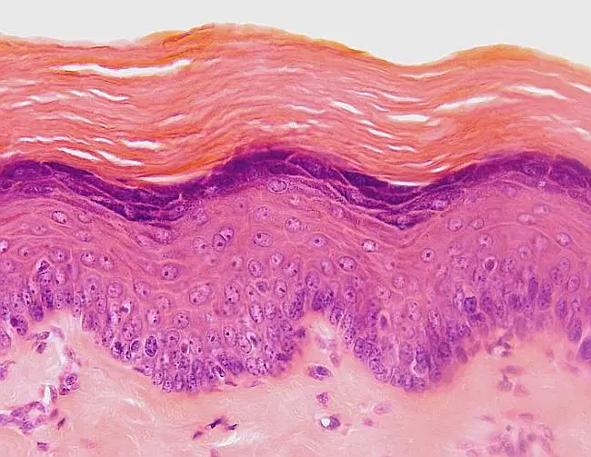
Keratinized Stratified Squamous
Pseudostratified Ciliated Columnar
Nonkeratinized Stratified Squamous
Simple Cuboidal
What type of tissue is this?
Muscle
Nervous
Epithelial
Connective

Stratified Squamous
Simple Squamous (top view)
Simple Columnar
Simple Cuboidal
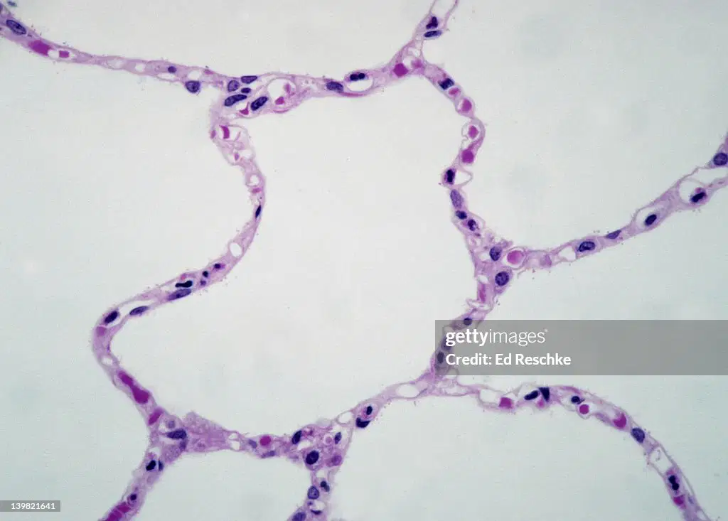
What tissue is this?
Stratified Squamous
Simple Squamous (side view)
Simple Columnar
Simple Cuboidal
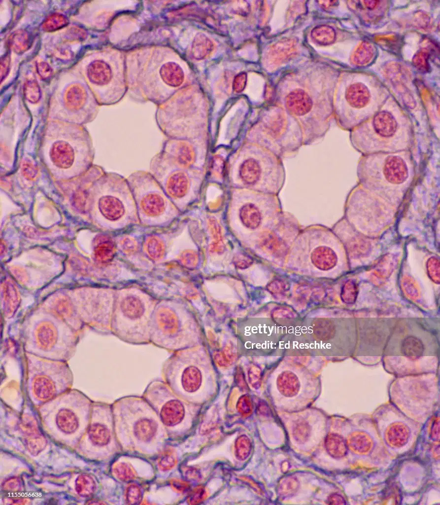
What tissue is this?
Simple Squamous
Pseudostratified Ciliated Columnar
Simple Columnar
Simple Cuboidal
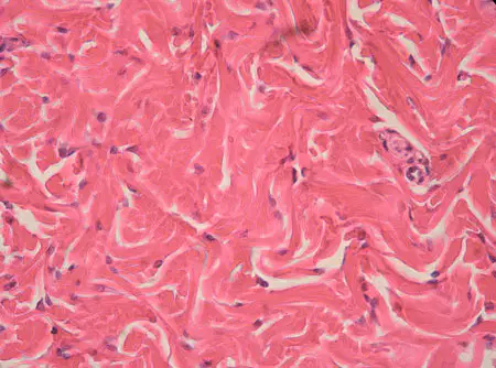
What tissue is this?
Dense Fibrous Regular
Hyaline Cartilage
Dense Fibrous Irregular
Bone
What type of tissue is this?
Muscle
Nervous
Epithelial
Connective
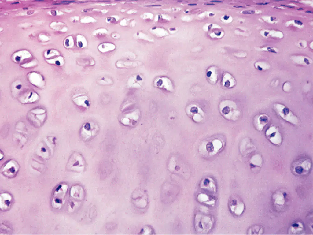
What tissue is this?
Hyaline Cartilage
Areolar
Blood
Bone
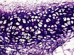
What tissue is this?
Hyaline Cartilage
Elastic Cartilage
Blood
Fibrocartilage

What tissue is this?
Keratinized Stratified Squamous
Pseudostratified Ciliated Columnar
Nonkeratinzed Stratified Squamous
Simple Cuboidal
What type of tissue is this?
Muscle
Nervous
Epithelial
Connective
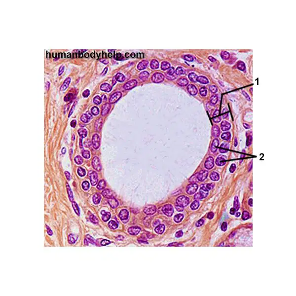
What tissue is this?
Stratified Cuboidal
Stratified Columnar
Simple Cuboidal
Simple Columnar
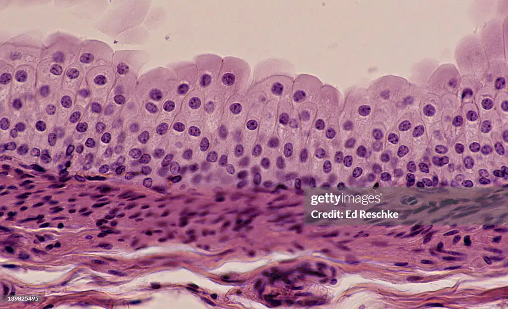
What tissue is this?
Stratified Cuboidal
Transitional
Simple Cuboidal
Simple Columnar
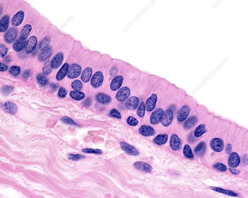
What tissue is this?
Stratified Cuboidal
Stratified Columnar
Simple Cuboidal
Simple Columnar
Define tissue with regards to anatomy
Multiple sheets of thin paper used to clean your nose
The smallest structural and functional unit of an organism
A series of different cells in the same area
A group of structurally and functionally related cells that come together to perform common functions
What is the difference between exocrine and endocrine functions?
Exocrine cells secrete fluids to the outside of the body; endocrine cells secrete fluids into the inside of the body
They are synonymous
Exocrine cells secrete fluids into ducts; endocrine cells secrete fluids into the blood stream
Exocrine cells secrete clear fluids; endocrine cells secrete opaque fluids

What landmark is labeled G?
olecranon notch
radial notch
styloid notch
trochlear notch
What landmark is labeled A?
Head of the Ulna
Head of the Radius
Neck of the Ulna
Neck of the Radius
What landmark is labeled M?
Styloid process of the radius
Styloid process of the ulna
Radial tuberosity
Ulnar tuberosity

What landmark is labeled 4?
greater tubercle
lesser tubercle
head
anatomical neck
What landmark is labeled 10?
medial epicondyle
lateral epicondyle
trochlea
capitulum
What landmark is labeled 1?
lesser tubercle
greater tubercle
deltoid tuberosity
radial tuberosity

Identify bone marking of the humeroulnar joint
trochlear notch
trochlea
olecranon fossa
olecranon process

Which of the following statements is correct.
this is the radius, it is located medially in the antebrachium
this is the radius, it is located laterally in the antebrachium
this is the ulna, it is located medially in the antebrachium
this is the ulna, it is located laterally in the antebrachium

Identify bone.
humerus
radius
ulna
fibula
tibia

Identify bones.
metacarpals
distal phalanges
proximal phalanges
middle phalanges
carpals

Identify bone marking #6
trochlea
trochlear notch
olecranon process
olecranon fossa
head of radius

Identify the landmark.
lateral malleolus, tibia
lateral malleolus, fibula
medial malleolus, tibia
medial malleolus, fibula
styloid process

Identify the highlighted region on the bone depicted above.
head of femur
neck of femur
greater trochanter
medial malleolus
patellar surface

Identify the highlighted region on the bone depicted above.
lesser trochanter
lateral condyle of femur
greater trochanter
linea aspera
medial condyle of femur

Identify this landmark
radial tuberosity
lesser tubercle
tibial tuberosity
femoral tuberosity
lesser trochanter

Identify this bone.
femur
patella
tibia
fibula
humerus

Identify the bone shown in green in the image:
talus
calcaneus
navicular
cuboid
cuneiform

Identify the bone shown in green in the image:
talus
calcaneus
navicular
cuboid
cuneiform

Identify the bones in green:
metatarsals
metacarpals
phalanges
tarsals
cuneiforms

Identify the bones in green:
metatarsals
metacarpals
phalanges
tarsals
cuneiforms

Bone #2 is...
Frontal Bone
Occipital Bone
Sphenoid Bone
Nasal Bone

Bone #3 is...
Parietal Bone
Zygomatic Bone
Sphenoid Bone
Maxilla

Bone #4 is...
Mandible
Occipital Bone
Maxilla
Temporal Bone

Bone #5 is...
Maxilla
Mandible
Frontal Bone
Occipital Bone

Bone #6 is...
Frontal Bone
Parietal Bone
Sphenoid Bone
Occipital Bone


What are the immovable joints between bones of the skull called?
foramen
joints
sutures
Structure A is the ________suture.
Coronal
Sagittal
Squamous
Lambdoidal

What is the name of the structure at the arrow?
External auditory (acoustic) meatus
Styloid Process
Mastoid Process
External Occipital Protuberance


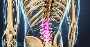
Which section of the vertebral column is shown?
cervical
lumbar
thoracic
sacral

Identify #4
Transverse process
Vertebral Body
Identify #3
Transverse process
Vertebral Body

Which section of the vertebrae is highlighted?
cervical
thoracic
lumbar
sacrum

What bone of the pelvic girdle is colored purple (#1)?
ilium
ischium
pubis
sacrum
coccyx
What bone of the pelvic girdle is colored pink (#3)?
ilium
ischium
pubis
sacrum
coccyx
What bone of the pelvic girdle is colored blue (#4)?
ilium
ischium
pubis
sacrum
coccyx
What bone of the pelvic girdle is colored blue (#5)?
ilium
ischium
pubis
sacrum
coccyx

Identify #16? It is the deep socket of the hip bone. It receives the head of the femur to form the ball and socket joint.
acetabulum
obturator foramen
iliac crest
greater sciatic notch
ischial spine
Identify #9? This projection is located on the posterior region. It allows for the attachment of various muscles and ligaments.
acetabulum
obturator foramen
iliac crest
greater sciatic notch
ischial spine

Identify the structure marked with the X: This cartilaginous joint joins the two pubic bones together.
pubic symphysis
obturator foramen
iliac crest
acetabulum

Which pelvis in the illustration is the male pelvis?
Pelvis A
Pelvis B
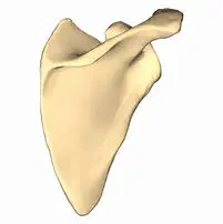
Name this bone.
Scapula
Clavicle
Spine
Glenoid Cavity
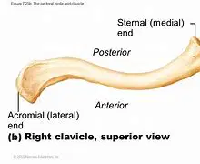
Name this bone.
Clavicle
Scapula
Spine
Glenoid Cavity
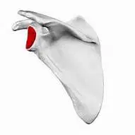
Name this structure.
Glenoid Cavity
Acromion Process
Spine
Suprascapular Notch

The muscular system interacts with the skeletal system by:
smooth muscles are joined with spongy bone via cartilage
skeletal muscles pull on joints to permit movement
cardiac muscle is connected to the ribs
skeletal muscles are joined to one another via ligaments
The sliding filament theory explains which of the following?
muscle extensions
muscle inductions
muscle contractions
muscle formation
______ and ______ work together to allow muscles to contract.
ATP, Calcium
smooth muscle, cardiac muscle
Electrolytes, Phosphorous
hemoglobin, plasma
Each unit in a muscle fiber is called a __________.
myosin
globular heads
sarcomeres
troponin
Which of the following is a way that the skeletal system is necessary for the muscular system's function?
the skeletal system has joints, which are what the muscular system pulls on to move
the skeletal system provides structure for the muscular system
the skeletal system provides calcium, which the muscles require along with ATP to contract
all of the above
Muscle used to grin
zygomaticus
buccinator
occiptialis
NONE
Muscle used to raise your eyebrows
frontalis
(frontal belly)
masseter
occipitalis
orbicularis oculi
This is the largest muscle of the buttocks
gluteus medius
gluteus maximus
gluteus minimus
NONE
This muscle allows you to extend your head
latissimus dorsi
trapezius
erector spinae
NONE
Triangle muscle that allows you to raise your arms laterally
deltoid
latissiums dorsi
trapezius
NONE
This muscle is used for blinking and squinting
buccinator
orbicularis oris
orbicularis oculi
zygomaticus
The prime mover in dorsiflexion.
Triceps brachii
Quadriceps
femoris
Biceps brachii
Tibialis anterior
The brachioradialis is the synergist to this muscle in flexion.
Triceps brachii
Quadriceps
femoris
Biceps brachii
Tibialis anterior
The prime mover for forearm extension
Biceps brachii
Triceps brachii
Quadriceps
femoris
Latissimus dorsi

Muscles attach to bone via:
ligaments
tendons

Within a sarcomere the thin myofilament is called:
Actin
Myosin

The connective tissue surrounding the muscle bundles is called:
Perimysium
Endomysium
Epimysium
Sarcolemma
What is the role of ATP in muscle contractions?
Regulates body temperature
Transmits nerve impulses to muscle fibers
Allows muscles to slide and prevent bursting
Provides energy for muscle contractions
The _________ separate one sarcomere from another?
synapses
m-lines
z-lines (z-discs)
active sites


Area #1 is called the -
Deltoid
Pectoralis major
Biceps Brachii
Brachialis

Area #2 is called the -
Deltoid
Pectoralis major
Biceps Brachii
Brachialis

Area #3 is called the -
Deltoid
Pectoralis major
Biceps Brachii
Brachialis

external oblique
intercostals
diaphragm
internal oblique

Identify the highlighted muscle
Deltoid
Biceps
Trapezius
Hamstring

Name this muscle
Hamstring
Quadricep
Gluteus Maximus
Rectus Abdominis

Name this muscle
Biceps brachii
Quadricep
Triceps brachii
Hamstring

Name this muscle
Pectorals
Latissimus Dorsi
Rectus Abdominis
Gastrocnemus

which skeletal muscle is represented in the image?
rectus femoris
vastus lateralis
vastus medialis
sartorius

#7
tibialis anterior
fibularis longus
soleus
gastrocnemius

#10 and #11
adductor group
abductor group
vastus medialis
iliopsoas

rectus femoris
vastus lateralis
vastus medialis
biceps femoris

This is what connects the gastrocnemius to the calcaneous bone (6)
gastrocnemius
calcaneous
achilles tendon
forman's ligament

Identify the muscle labeled R
hamstrings
gluteus medius
quadriceps
fibularis

Identify this muscle
hamstrings
quadriceps
gastronecmius
sartorius

This muscle crosses the legs over one another (8)
sartorius
hamstrings
Forman's crossorius
extensor carpi radialis

