Explore Year 9 skull bones through Wayground's comprehensive biology worksheets featuring printable PDFs, practice problems, and answer keys to help students master cranial and facial bone identification and functions.
Explore printable Skull Bones worksheets for Year 9
Year 9 skull bones worksheets available through Wayground (formerly Quizizz) provide comprehensive anatomical study materials that help students master the complex structure of the human cranium and facial skeleton. These educational resources focus on identifying and understanding the twenty-two bones that comprise the skull, including the frontal, parietal, temporal, and occipital bones of the cranium, as well as the mandible, maxilla, nasal, and zygomatic bones of the facial region. The worksheets strengthen critical skills in anatomical terminology, spatial reasoning, and memorization techniques through varied practice problems that range from labeling diagrams to matching bone names with their functions. Students benefit from structured learning materials that include detailed answer keys, ensuring accurate self-assessment and reinforcement of correct anatomical knowledge. These free printables serve as invaluable study aids that complement textbook learning and prepare students for assessments in human anatomy and biology.
Wayground (formerly Quizizz) supports educators with an extensive collection of skull bones worksheets created by millions of teachers worldwide, offering unparalleled variety and quality in anatomical education resources. The platform's robust search and filtering capabilities enable teachers to quickly locate materials that align with specific curriculum standards and match their students' learning needs. Advanced differentiation tools allow educators to customize worksheets for diverse learners, from struggling students requiring additional visual supports to advanced learners ready for more complex anatomical relationships. These resources are available in both printable pdf formats for traditional classroom use and digital formats for interactive learning environments, providing maximum flexibility for lesson planning and implementation. Teachers utilize these comprehensive materials for initial skill instruction, targeted remediation sessions, enrichment activities for accelerated students, and regular practice opportunities that reinforce proper identification and understanding of skull bone structures throughout their Year 9 biology curriculum.
FAQs
How do I teach skull bones to students effectively?
Teaching skull bones is most effective when students first understand the skull's two major divisions: the cranium (which protects the brain) and the facial skeleton (which supports sensory organs and structures like the jaw). Begin with the eight cranial bones before introducing the 14 facial bones, using labeled diagrams to anchor each structure spatially. Connecting each bone to its function, such as how the temporal bone houses the auditory canal or how the occipital bone surrounds the foramen magnum, helps students retain names through meaningful context rather than rote memorization.
What exercises help students practice identifying skull bones?
Labeling diagrams is the most direct practice method for skull bone identification, requiring students to place bone names on unlabeled lateral, anterior, and superior views of the skull. Suture-mapping exercises, where students trace and name the coronal, sagittal, lambdoid, and squamous sutures, reinforce spatial relationships between bones. Matching functions to structures, such as pairing the mandible with chewing or the nasal bone with the bridge of the nose, builds applied anatomical understanding beyond simple recall.
What mistakes do students commonly make when learning skull bones?
Students frequently confuse the temporal and parietal bones because both form lateral portions of the cranium and share a border along the squamous suture. Another common error is misidentifying the sphenoid bone, which is not visible on a standard lateral view and is often omitted from student answers entirely. Students also tend to conflate the maxilla and mandible, incorrectly treating both as movable, when only the mandible articulates at the temporomandibular joint.
How can I differentiate skull bones instruction for students with different learning needs?
For students who struggle with dense anatomical vocabulary, reducing the number of bones targeted in a single session and focusing on major cranial bones before facial bones can lower cognitive load. On Wayground, teachers can enable Read Aloud so bone names and questions are read to students who benefit from audio support, and can apply reduced answer choices for students who need a more guided identification task. Extended time settings can also be assigned to individual students without affecting the rest of the class, making it straightforward to accommodate diverse learners during digital assessments.
How do I use Wayground's skull bones worksheets in my classroom?
Wayground's skull bones worksheets are available as free printable PDF downloads for paper-based anatomy lessons and in digital formats for technology-integrated classrooms. Teachers can assign them as independent practice, homework, or structured review activities. The digital versions can also be hosted as a quiz on Wayground, giving teachers real-time insight into student performance on specific bone identification tasks. Each worksheet includes a detailed answer key, so grading and self-checking are built in.
How many bones make up the human skull, and which ones do students need to know?
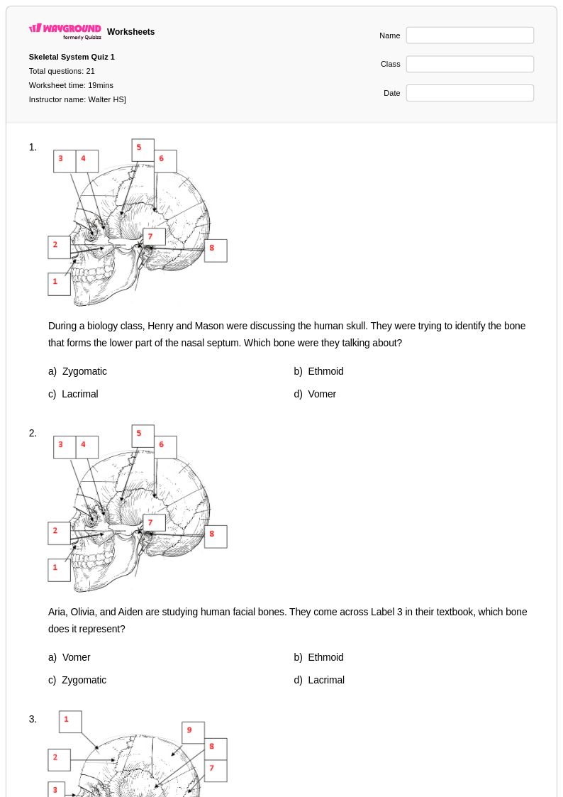
The human skull is composed of 22 bones in total: 8 cranial bones and 14 facial bones. The cranial bones students most commonly need to identify include the frontal, parietal (paired), temporal (paired), occipital, sphenoid, and ethmoid. Key facial bones include the nasal, maxilla, mandible, zygomatic, lacrimal, and palatine bones. Most secondary anatomy courses prioritize the cranial bones and the larger facial bones, while more advanced coursework extends to smaller structures like the vomer and inferior nasal conchae.